Introduction
Skin, the largest organ in the human body, plays an important role of providing mechanical and immune defense against environmental insults, including microorganisms, trauma, and environmental stress (1). Disruption of skin integrity resulting from trauma, burns, surgical procedures, diabetes, or vascular disease could cause tissue damage, either acute or chronic (2). Acute wounds normally complete the reconstruction process and restore tissue integrity in a matter of time, whereas chronic wounds, (e.g., Diabetic foot ulcers, venous leg ulcers, and pressure sores) show reduced or abnormal healing with intense inflammation, infection, deficient angiogenesis, and abnormalities of basic metabolic regulation (3). These conditions generally result in prolonged treatment, higher cost of healthcare, worsened quality of life, and more chances for complications, which is why there is a necessity for new and effective ways of treatment. Skin regeneration is a very complicated and organized biological sequence during which four phases—hemostasis, inflammation, proliferation, and remodeling—take place overlapping each other (4). At the hemostasis stage blood clotting hinders further bleeding as well as functioning. They act as a temporary scaffold for cell, migration, then at the inflammation stage immune cells are recruited to the wound to kill bacteria and clear dead cells (5). The proliferation phase is characterized by the multiplication of fibroblasts and the production of the interstitial matrix [extracellular matrix (ECM)]; also, new capillaries are formed, and the epithelium is using epidermal cells to cover the surface. In the final phase of tissue remodeling, the newly formed tissue is getting stronger and stronger via collagen maturing and the reorganization of tissue (6). Fibroblast communication and cytokine signaling, controlled inflammation, proper blood supply, and ECM turnover should always work in harmony in the wound microenvironment for the tissue to regenerate successfully (7). From this point of view, scaffold-assisted regenerative therapies have become a good alternative to traditional wound dressings because, apart from physically supporting the wound healing process, they are also able to help cell growth and act as a vehicle for therapeutic agent delivery.
Scaffold fabrication methods are concerned; the electrospinning technique stands out as an amazing method to produce extremely thin fibers that are similar in size to the fibrous elements of the native ECM (8). Besides having the properties of ECM in a physical sense, electrospun nanofibers are made of interconnected porous structures, have a high surface-area-to-volume ratio, their mechanical properties can be adjusted, and they have excellent oxygen permeability and supply of nutrients making them one of the best materials for skin tissue engineering (9). Their morphology, which mimics the ECM, greatly increases the performance of fibroblasts through their enhanced attachment, keratinocyte migration, collagen production, and the formation of new vessels (10). Besides that, these nanofibers keep the wound in a moist environment, which is one of the main factors that contributes to a rapid healing of such wounds. And electrospun nanofibers are a handy tool to deliver different bioactive components, including antimicrobial agents, different growth factors, anti-inflammatory agents, and various nanoparticles, which allow multifunctional wound dressings with regulated therapeutic release.
In the field of wound care, many types of nanomaterials have been used, but among all of them silver nanoparticles (AgNPs) stand out the most. Silver is known to have an excellent antimicrobial effect not only against common Gram-positive and Gram-negative bacteria but also against fungi and various strains resistant to antibiotics, which are the ones that lead to wound infections (11). AgNPs’ effectiveness against microbes consists of destruction of the microbial cell wall with massive leakage of cell constituents, production of reactive oxygen species (ROS), hindermost of the microbial enzymes, inhibition of DNA replication, and blockage of biofilm formation (12). Apart from their antimicrobial effects, AgNPs also possess anti-inflammatory, antioxidant, pro-healing properties that, in all, make the process of wound healing faster (13). AgNPs assist in wound healing by altering inflammatory mediators, boosting skin cell growth and migration, causing new blood vessel formation, and increasing collagen accumulation. Yet, deciding on the best concentration of silver and the way it is released are two important issues that should be addressed so that not only the therapeutic effect is maximized but also the toxicity to human cells is also minimized.
Integrating AgNPs into electrospun nanofibrous scaffolds is a very effective way to make wound dressings that not only provide structural support but also have sustained antimicrobial and cell-regenerative properties. These composites offer targeted silver release, decrease the chances of bacterial infection, improve the interaction of cells with the biomaterial, and help the skin to heal more rapidly. This mini review highlights the progress made in electrospun nanofibers incorporating AgNPs for skin tissue regeneration.
Electrospun nanofibers as skin tissue engineering scaffolds
Electrospinning is an easy and highly adaptable method to make nanofibrous scaffolds that have a similar structure to the natural ECM of skin cells (14). It involves the use of a very high voltage on a polymer solution or melted polymer material and throwing this fine jet of polymer particles in the air. With the evaporation of solvent, they get hard and continuous nanofibers. Finally, the fibers are collected in the form of a porous, nonwoven mat with modifiable morphology and mechanical properties. By changing different processing parameters like polymer concentration, voltage applied, flow rate, and distance between the collector and the needle, the electrospun scaffolds can be designed to completely fulfill targeted skin tissue engineering needs (15).
Electrospun nanofibers have a number of benefits over traditional wound dressings like gauze and hydrocolloids, which help skin regeneration. Firstly, the fibrous structure almost resembles the natural ECM, providing an ideal platform for cell activities like attachment, growth, and movement. Secondly, through their high porosity and interconnected pore structure, the exchange of nutrients and removal of wastes as well as the vascularization process are enhanced, whereas the high surface-area-to-volume ratio is conducive to the adsorption of proteins and subsequent cell-material interactions (16). Also, electrospun nanofibers are highly permeable to oxygen and retain moisture very well, which keeps the wound environment at an optimal level to support re-epithelialization and lower the risk for infection (17). Due to their very high capacity for drug loading, substances like antimicrobial agents, growth factors, nanoparticles, and other bioactive molecules can be incorporated for sustained and localized therapeutic release to promote wound healing.
The polymer type can greatly impact the biological and the mechanical properties of electrospun scaffolds. Natural polysaccharides, proteins, and other biopolymers like chitosan, gelatin, collagen, silk fibroin, and alginate are the first choices of researchers for making such scaffolds due to their excellent biocompatibility, biodegradability, and the ability of cells to adhere, survive, and multiply on them (18). Besides, chitosan contains antimicrobial and hemostatic agents, collagen, and gelatin are best to mimic ECM for cell attachment; silk fibroin is a very strong material and elicits little immune response; and alginate can hold a lot of water and forms gels, making it suitable for highly exuding wound situations.
Synthetic polymers are added to natural ones for improving the mechanical properties and stability of the scaffold (19). Polycaprolactone (PCL) stays one of the most popular polymers, being highly flexible, biocompatible, biodegradable, and having a slow degradation rate (20). Polyvinyl alcohol (PVA), without a doubt, stands very high in electrospinnability and hydrophilicity, and poly(lactic acid) (PLA) and poly(lactic-co-glycolic acid) (PLGA) are capable of providing mechanical support and controlled biodegradation (21–23). Another polymer, polyurethane (PU), is well-known for its elasticity and durability. PU-based wound dressings can be stretched and bent repeatedly without changing their structures (24).
Silver nanoparticles in skin tissue regeneration
Ag NPs are becoming increasingly popular for skin tissue engineering due to their special physicochemical properties and versatile biological activities. Their nanoscale size, large surface-area-to-volume ratio, and high surface reactivity are proven to enable Ag NPs to have a greater interaction with microbial cells and biological tissues than bulk silver (25). Features like particle size, shape, surface charge, and surface functionalization affect the physicochemical properties of AgNPs, which in turn determine their performance with stability, antimicrobial effect, cellular uptake, and biocompatibility (26). Smaller particles generally offer higher antimicrobial activity because they expose a larger reactive surface area, but a suitable surface modification may also improve dispersion, prevent aggregation, and enable the controlled release of silver ions. These characteristics of AgNPs make them very attractive materials for the production of wound healing dressings and tissue engineering scaffolds.
Besides being antimicrobial, which effectively kills the most variety of bacteria, fungi, and even those pathogenic drug-resistant microbes (27). AgNPs have multiple wound-healing factors, which include killing micro-organisms in various ways at once, which practically stops micro-organisms from developing resistance. First, they adsorb to and penetrate the microbial cell membrane, thereby causing higher membrane permeability and cellular content leakage through the membrane. Following entering the cells, AgNPs generate ROS, which through oxidative stress, leads to the destruction of important cell parts like the DNA. It bind to the DNA of the microbe, hindering DNA replication and division of cells; simultaneously, it will attach to the microorganism’s proteins and enzymes, resulting in protein denaturation or breakdown of the enzymes and the prevention of vital metabolic pathways (28). AgNPs are also very strong inhibitors of the formation and maturation of microbial biofilms, which are the major cause of chronic infections and the wound healing delay. Using AgNPs to hinder biofilm development will result in better infection control and create a more appropriate environment for tissue regeneration.
Apart from their antimicrobial effects, AgNPs also participate in various biological functions that are vital for tissue repair and regeneration (29). For instance, by reducing excessive inflammatory reactions and limiting the release of pro-chemicals from the cell surface, anti-inflammatory functions prevent tissue degradation in a non-healing wound (30). This mild inflammatory situation helps the change from the inflammatory to the proliferative phase of wound healing. Also, it was discovered that AgNPs can improve angiogenesis through the development of new blood vessels, a process that is vital for supplying fresh oxygen and nourishment to the regenerating tissues (31). This enhancement in vascularization allows the wound bed to be covered by granulated tissue more quickly and nourish the skin cells living at the site.
AgNPs also have a positive effect on increasing the migration and proliferation of fibroblasts, cells that regulate different processes on ECM and tissue contraction (29). This increase in activity of fibroblasts results indirectly in formation of more collagen, a vital component providing structural integrity and tensile strength to the newly formed tissue (Figure 1). In addition, AgNPs assist epidermal cells or keratinocytes, to migrate and multiply, a key process for the healing of epithelium or re-establishment of an epidermal barrier layer (32). All these properties of AgNPs have brought about faster healing and a decrease in the rate of infection, besides tissue architecture restoration and, finally, functional recovery.
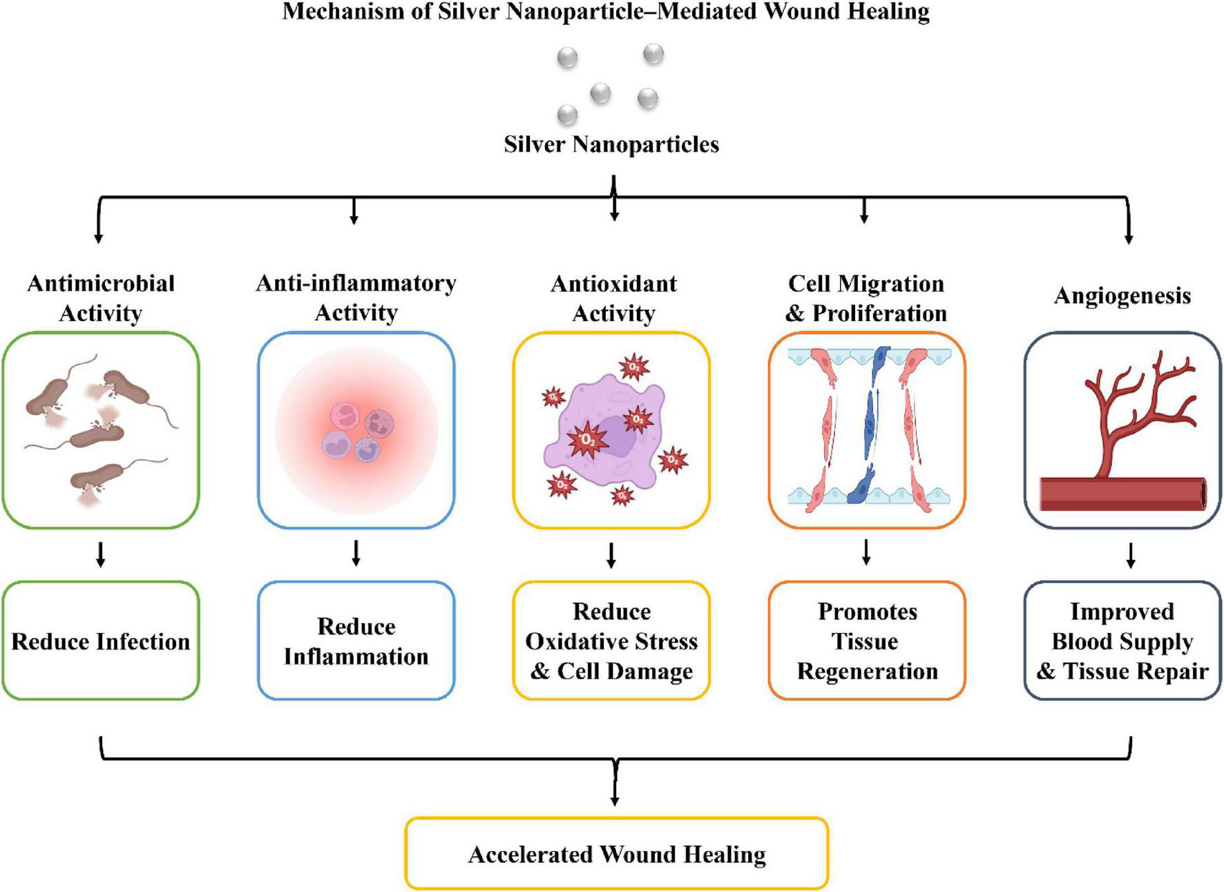
Figure 1. Mechanism of AgNP-mediated wound healing.
Fabrication of silver nanoparticle- loaded electrospun nanofibers
Fabricating nanofiber wound dressings with antibacterial properties and healing-promoting qualities for a long time can be effectively achieved with the electrospun AgNP nanofibrous approach (33). Through different routes, these AgNP-containing wound dressings have been designed to ensure nanoparticles are uniformly scattered, silver is discharged in a gradual manner, and the dressings have biocompatible characteristics. Blending electrospinning technique is the simplest and most widespread among those routes: AgNPs prepared earlier were directly added to the polymeric solvent, which is then used for electrospinning. The procedure proved to be effective for even distribution of the nanoparticles within nanofibers and very easy to carry out (34). Though, improper optimization of the dispersion could result in agglomeration of nanoparticles and a large burst release of silver ions during the initial stages.
Surface immobilization is a different method often used where AgNPs are fixed to the surface of the electrospun nanofibers that have been made earlier via physical adsorption or chemical bonding. Since the nanoparticles stay on the fiber surface, this is a way of giving microbes more direct access to them, which can cause better antimicrobial effectiveness in some cases. Then again, if the surface interactions are weak, the nanoparticles may come off during use for a longer period of time. In situ synthesis is an alternative method that is becoming more popular where silver ions are first added to the electrospun scaffold, and then they are changed into AgNPs either inside or on the fibers (35). This approach usually results in nanoparticles that are very well spread out and strongly bonded to the polymer matrix, which stabilizes the nanoparticles and also reduces aggregation.
Electrospinning methods that are more sophisticated have made it possible to develop more designs for wound dressings containing AgNPs. With coaxial electrospinning, two nozzles, one inside the other, are used. This way, it is possible to make core-shell nanofibers so that AgNPs can be put only in the core or the shell layer (36, 37). This design keeps the nanoparticles safe and makes it possible to release silver slowly and in a controlled way, which reduces potential cell damage due to rapid ion release. Also, emulsion electrospinning uses polymer solutions that have been emulsified to make fibers that have core-shell-like structures in a single-step procedure. Through this method, it is possible to encapsulate AgNPs and other therapeutic agents simultaneously, which makes it really very useful for the development of multifunctional wound dressings that combine antimicrobial, anti-inflammatory, or regenerative activities.
The work of AgNPs-containing electrospun scaffolds is affected by a number of factors related to fabrication. Fiber size or diameter plays a big role because it affects the surface area, mechanical properties, degradation behavior, and release of silver. Nanofibers that are smaller in diameter tend to have a bigger surface area, which enables better cell attachment and offers more amount of antimicrobial agents (38). Besides that, another very essential parameter is the loading or concentration of AgNPs. A smaller amount may result in not very good performance against microorganisms. Yet, an excessive amount of silver may be toxic to the cells and also prevent the development of new tissues. That means determining the amount of AgNPs is extremely crucial to combine the properties of effective antimicrobial and compatibility with the body.
Therapeutic applications in skin tissue regeneration
AgNP-loaded electrospun nanofibers are one of the most effective biomaterials for skin regeneration owing to the combined advantages of both nanofibrous scaffolds and the versatile role of AgNPs as microbial fighters and tissue regrowth. Because of their similarity with natural ECM, they can not only support cell functions that are needed for tissue growth but also, through the silver ion charge, are a source of protection from microbial infections. Their super abilities have led to their deployment in smart wound dressing solutions for skin damage caused by both sudden and long-term conditions.
AgNP-loaded electrospun nanofibers resist a wide spectrum of microbes, which is the cornerstone of their appearance in antibacterial wound dressings. Besides interfering with the bacterial proliferation and biofilm formation, the continuous silver ion release from nanofibers containing AgNPs is capable of reducing the colonization of microbes at the wound location, which in itself is a highly positive point for wound healing (39). The exchange of oxygen and the moisture level maintaining by the fiber’s porousness help not only in tissue regeneration but also in putting the patient’s life on the least-used dressing track.
AgNP-loaded electrospun materials are an effective weapon against the microbial challenge in burn wounds, which often suffer from losing large amounts of fluids besides the physical injury to the skin barrier. Making the infection control part of the burn procedure, entry of Pseudomonas infection to the damaged tissue, attracting inflammatory cells for hours, and late necrosis of new skin (40). Having AgNPs in the burn dressing can help to not only fight all these aspects of infection but can also be involved in the process of reducing the wound’s inflammation level, safeguarding the wound with a moist environment, and supporting fibroblast growth, collagen networking, sprouting of new vessels, etc., all taking the skin above the baseline toward full recovery. Table 1 summarize the AgNP-loaded nanofibers for wound healing applications.
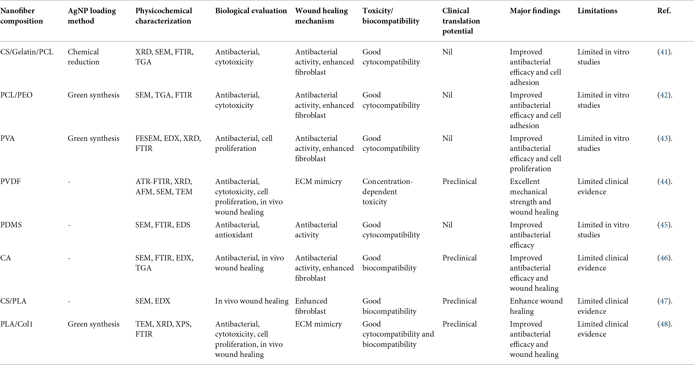
Table 1. Comparative summary of AgNP-loaded nanofibers for wound healing applications.
Among the different fabrication strategies of AgNPs-loaded electrospun nanofibers, green synthesis and in situ chemical reduction methods have been identified as highly effective because they not only allow for uniform distribution of nanoparticles but also reduce the use of harmful reducing agents. Green synthesis is the main principle behind those systems like PCL/PEO, PVA, and PLA/Col1 nanofibers, which have been consistently found to have the highest antibacterial effect, promote fibroblast proliferation, and have excellent cytocompatibility. So, they seem to be the best suited for wound healing (42, 43, 48). Mainly, the PLA/Col1 scaffold prepared by green synthesis combined antibacterial properties with ECM mimics, cell proliferation, and wound healing in vivo, which showed the most biocompatible features (48). On the opposing front, the chemically reduced CS/Gelatin/PCL nanofibers presented quite well for killing bacteria and allowing fibroblasts to stick; yet, only their in vitro test results were presented, and because of this, their therapeutic efficacy for the long run remained uncertain (41). Besides, some studies described AgNPs without giving details of the loading method; still, electrospun PVDF and cellulose acetate (CA) nanofibers showed exceptional wound healing results in animal experiments due to their structural integrity, persistent antibacterial activity, and tissue regeneration (44, 46). Overall, available data indicate that green synthesis coupled with electrospinning offers the optimal compromise between biological properties, nanoparticle stability, and environmental friendliness; yet further detailed comparative studies of various AgNP loading methods all with the same experimental conditions are still needed.
The biological and mechanical characteristics of biocomposite wound dressings are largely dependent on the type of polymer used as the matrix. For example, PVDF not only gives excellent mechanical resistance but also makes antibacterial activity, cell proliferation, and healing acceleration in vivo possible simultaneously, which is why PVDF became one of the most promising wound dressings with the highest mechanical strength (44). Its dosing level, though, has been found to be cytotoxic, dependent on concentration, which calls for the fine-tuning of AgNP dosage (44). A natural-polymer-based composite like PLA with collagen type 1 or CS with gelatin/PCL is expected to have better biological functioning in that they closely match the composition of a native ECM; it allows for improvement of fibroblast adhesion, proliferation, and tissue regeneration without affecting cell compatibility positively (41, 48). CA nanofibers in the same line of argument showed high biocompatibility tied to very significant antimicrobial activity and improved healing of wounds in preclinical evaluations (46). Chitosan/PLA composites, in their turn, contributed to the tissue regeneration due to reality, chitosan is bioactive, and they presented an encouraging result on the wound healing of animals (47). But if PCL/PEO and PVA have the advantage of antibacterial activity and promoting cell proliferation, then the mechanical performance and the long-term behavior after implanting these scaffolds need further investigations (42, 43). Overall, the research results support that the strategy of a dual polymer system in which the artificial polymer gives the mechanical properties while the biological part is provided by the natural polymer for cell interaction is the way to go if one wants a wound dressing based on electrospun fibers loaded with AgNP capable of both structural and biological functions (41, 44, 46–48).
Challenges and future perspectives
One reason why a broad adoption of Ag NP-loaded electrospun nanofibers is being slowed down, besides the promising regenerative effect on skin tissue that they may produce, is the existence of some issues that need to be resolved. One of the main challenges is about the toxic effect of silver. A high dose of silver ions or an uncontrolled release of a very large amount of these ions might lead to oxidative stress, reduction of the viability of various skin cells, and delay in tissue regeneration. So, the nanoparticles’ size, concentration, and distribution should be highly adjusted for achieving a suitable equilibrium between the antibacterial potential and the cell compatibility. Besides that, the so-called burst release, the release of silver ions instantly following the scaffold’s introduction into the body, could bring a short-lived antimicrobial effect while a bit increasing the risk of damage to cells. Research on nanomaterial-based dressings is focused mainly at present on the fabrication of silver-releasing scaffolds, which, unlike the former, can deliver silver ions in a continuous and safe manner. Though, the long-term biocompatibility of AgNP-containing matrices remains a question. While many lab and animal models have shown successful outcomes of wound healing, only a limited number of detailed, comprehensive investigations with nanoparticle accumulation, degradation behavior, and systemic safety have been published. Also, lack of a standardized way to make the product causes inconsistencies when it comes to getting the fibers’ shape, nanoparticles’ dispersion, mechanical performance, and the therapeutic results of the samples made from different research groups. Variations in the electrospinning parameters, the type of polymers and the methods of nanoparticles synthesis are commonly the main factors why one sample is not comparable to another with direct comparison and why the results cannot easily be reproduced.
Scaling a technology or method from a laboratory level to commercial manufacturing level often presents additional challenges. The fact is, electrospun nanofibers, which still maintain their quality about uniformity, reproducibility, and cost-efficiency, can even so be manufactured on an industrial scale only if these technical issues are first resolved. But, besides that, consider that before the regulatory authorities allow nanotechnology-based wound dressings to be marketed, the manufacturers are going to need proof of safety through extensive preclinical studies, clinical trials, and compliance with strict manufacturing standards. All this will probably lead into a significant increase of development time as well as development costs. Though it is a rather straightforward operation, at the laboratory level, the large-volume manufacturing of electrospun nanofibers, which also takes into account high-quality polymers, nanoparticle synthesis, sterilization, and quality control, is the cost-efficiency issue that can really put off the use in a clinical setting. New research might take a path of developing highly effective electrospun wound dressings for practical applications. Besides aiding optimization of fiber architecture, polymer composition, and nanoparticle loading using predictive scaffold performance and therapeutic outcomes, artificial intelligence (AI)-assisted scaffold designing may usher in a new era of wound dressings. Everyone knows that diabetic wounds tend to become chronic ones, and a dressing personalized to a patient’s unique characteristics and conditions of the wound would be highly effective. Controlled-release nanofibers that have advanced for a long time are expected to work as delivery systems of silver ions that are both sustained and have least toxicity and yet have an antimicrobial effect for a long time.
Though preclinical data is promising, there are a few main reasons that the development of AgNP-loaded electrospun nanofibers has not made it to real-life medical applications yet. The major problem is that there is almost no clinical evidence—all research has really been limited to studies in tubes or test animals. Only some of these systems, like PVDF CA CS/PLA and PLA/Col1 nanofibers, got validated through preclinical studies (44, 46–48). Also, another big reason is that releasing too much silver ions can be harmful to cells, mostly for those cases of PVDF where studies show that higher concentrations are actually toxic (44). Besides, many studies have only looked into antibacterial and cytocompatible activities and have ignored biodegradation, pharmacokinetics, systemic silver accumulation, immunological responses, or chronic toxicity (41–48). There are also no uniform electrospinning standards or AgNP loading procedures that would allow consistent release of AgNP and reliable manufacturing. Limited evaluation of stability on a shelf, sterilization ability, compatibility with storage, and cost-effective production methods also hinder the move towards commercialization. Because of this, although AgNP-loaded electrospun nanofibers possess a considerable potential, only clinical translation that includes standardized methods of production, in-depth biosafety evaluations, industrial manufacturing techniques, and carefully planned out clinical trials may result in successful results (44, 46–48).
There are many crucial topics left to explore in the creation of AgNP-loaded electrospun nanofibers for wounds. A large majority of the available research is focused on antibacterial properties and very short time period cytocompatibility with few explanations about the molecular mechanisms for the processes involved in healing such as angiogenesis, collagen inflammation, polarization of macrophages, and growth factors during wound repair (41, 48). Comparative studies of different polymer matrices, AgNPs synthesis methods, and loading techniques under similar experimental conditions that would help to pinpoint the best scaffold design are not performed either. Besides, there haven’t been many research efforts to optimize the levels of AgNP that, on one hand, deliver the best treatment effect and, on the other hand, the least cell toxicity because of silver, even though there were some accounts of concentration-dependent toxicity in some setups like PVDF (44). The way these dressings degrade over time and the release of silver at the right rate as well as how they interact with a wound microenvironment and their potential of these dressings in fighting against polymicrobial and antibiotic-resistant infections is an area that needs more attention and study. Most importantly, there is a great lack of translation research through large animal models, GMP-compatible fabrication processes, regulatory evaluations, and randomized clinical trials (44, 46, 48). Bridging the described shortcomings by involving experts across the fields of materials science, nanotechnology, molecular biology, and clinical investigation shall For sure, it will be the key for moving AgNP-loaded electrospun nanofibers from prototypes in labs to clinical wound care products. It will also be necessary to do multidisciplinary investigations if we hope for the latter to become a reality.
Though, there must be a lot of work done in the field of clinical translation of such types of wound dressings involving thorough preclinical studies, large-scale clinical trials, and the establishment of standardized manufacturing protocols to help with the regulatory approval process. In the near term, the new wound dressings may include AgNPs combined with growth factors, stem cells, antimicrobial peptides, anti-inflammatory agents, or natural bioactive compounds to obtain therapeutic synergy, and this way, interaction between the various therapeutic agents will be essential. The appearance of multi-functional bioactive scaffolds that can do what comes next simultaneously, avert infection, resolve inflammation, encourage angiogenesis, and facilitate tissue regeneration will be a promising development in the forthcoming generation of wound care products. Introducing a different point of view that collaborating across disciplinary borders should be a critical element in addressing these problems, alongside working between materials scientists, biomedical engineers, clinicians, and the regulatory agencies. This will make it possible to change AgNP-loaded electrospun nanofibers from being just a research item to becoming a routine clinical practice.
Conclusion
Electrospun nanofibers are set to become the new generation of scaffolds for skin tissue engineering since their architecture, which mimics ECM, generates a suitable microenvironment that can support cell adhesion, proliferation, and tissue regeneration. Adding AgNPs to these scaffolds has increased their efficiency as therapeutic agents by providing them with features like broad-spectrum antimicrobial, anti-inflammatory, and regenerative properties that together speed up wound healing, and at the same time, the risk of infection is minimized. However, the release of silver in a controlled and sustained manner is paramount for enhancing therapeutic effects and, at the same time, maintaining cytocompatibility through the reduction of the likelihood of side effects caused by excessive exposure to silver. Smart and multifunctional electrospun nanofiber systems that are responsive to stimuli and include bioactive scaffolds have recently been introduced, and these can be used for the treatment of chronic and infected wounds effectively. Although the outcomes of pre-clinical studies are very encouraging, there is still a need for additional long-term safety studies, suitably designed clinical trials, methods of fabrication that are standardized, and manufacturing processes that can be scaled up to speed up the translation of these advanced wound dressings into everyday clinical practice. Generally, electrospun nanofibers loaded with AgNPs constitute a very attractive platform for skin tissue regeneration and wound management in the future.
Ethics approval
Not applicable.
Data availability statement
No new data were created or analyzed in this study.
Funding
The author declares that financial support was not received for this work and/or its publication.
Conflict of interest
The author declares that the research was conducted in the absence of any commercial or financial relationships that could be construed as a potential conflict of interest.
References
2. Levine JM. Pressure injury and chronic wounds. In: Wasserman MR, Bakerjian D, Linnebur S, Brangman S, Cesari M, Rosen S editors. Geriatric Medicine: A Person Centered Evidence Based Approach. Cham: Springer (2024). p. 1185–212.
3. Jeyaraman N, Roy M, Muthu S, Khan S, Jeyaraman M. Preclinical insights into wound healing: models, mechanisms, and translational strategies. In: De Francesco F, Turksen K editors. Management and Strategies for Wound Healing. Springer (2026). p. 287–309.
4. Peña OA, Martin P. Cellular and molecular mechanisms of skin wound healing. Nat Rev Mol Cell Biol. (2024) 25(8):599–616.
5. Priya SES, Taj T, Fakrudheen F, Sahana TG, Ahmed MG. Introduction to wound healing: factors affecting, phases, and principles of wound healing. In: Ahmed MG, Ashique S, Farid A, Zengin G editors. Nanomaterials for Wound Healing. Boca Raton (FL): CRC Press (2025). p. 1–18.
6. Hamze J, Broadwin M, Stone C, Muir KC, Sellke FW, Abid MR. Developments in extracellular matrix-based angiogenesis therapy for ischemic heart disease: a review of current strategies, methodologies and future directions. BioTech (Basel). (2025) 14(1):23.
7. Huang J, Heng S, Zhang W, Liu Y, Xia T, Ji C , et al. Dermal extracellular matrix molecules in skin development, homeostasis, wound regeneration and diseases. Semin Cell Dev Biol. (2022) 128:137–44.
8. Adel IM, ElMeligy MF, Elkasabgy NA. Conventional and recent trends of scaffolds fabrication: a superior mode for tissue engineering. Pharmaceutics. (2022) 14(2):306.
9. Chamanehpour E, Thouti S, Rubahn HG, Dolatshahi-Pirouz A, Mishra YK. Smart nanofibers: synthesis, properties, and scopes in future advanced technologies. Adv Mater Technol. (2024) 9(3):2301392.
10. Hama R, Reinhardt JW, Ulziibayar A, Watanabe T, Kelly J, Shinoka T. Recent tissue engineering approaches to mimicking the extracellular matrix structure for skin regeneration. Biomimetics (Basel). (2023) 8(1):130.
11. More PR, Pandit S, Filippis A, Franci G, Mijakovic I, Galdiero M. Silver nanoparticles: bactericidal and mechanistic approach against drug resistant pathogens. Microorganisms. (2023) 11(2):369.
12. Parmar S, Kaur H, Singh J, Matharu AS, Ramakrishna S, Bechelany M. Recent advances in green synthesis of Ag NPs for extenuating antimicrobial resistance. Nanomaterials (Basel). (2022) 12(7):1115.
13. Balaure PC, Niculescu A, Anghel D, Grumezescu AM, Alberts A. Role of silver nanoparticles in wound healing: mechanisms, efficacy, and clinical applications. Inorganics. (2025) 13(12):401.
14. Wu J, Yu F, Shao M, Zhang T, Lu W, Chen X , et al. Electrospun nanofiber scaffold for skin tissue engineering: a review. ACS Appl Bio Mater. (2024) 7(6):3556–67.
15. Yu H, Tan Z. Electrospinning basics. In: Introduction to Electrospinning and Nanofiber. Cham: Springer (2025). p. 7–37.
16. Zhang X, Wang Y, Gao Z, Mao X, Cheng J, Huang L , et al. Advances in wound dressing based on electrospinning nanofibers. J Appl Polym Sci. (2024) 141(1):e54746.
17. da Costa PRA, de Menezes LR, Dias ML, da Silva EO. Advances in the use of electrospinning as a promising technique for obtaining nanofibers to guide epithelial wound healing in diabetics—mini-review. Poly Adv Technol. (2022) 33(4):1031–46.
18. Mahmud MZA, Islam MD, Mobarak MH. The development of eco-friendly biopolymers for use in tissue engineering and drug delivery. J Nanomater. (2023) 2023(1):1–15.
19. Keshvardoostchokami M, Majidi SS, Huo P, Ramachandran R, Chen M, Liu B. Electrospun nanofibers of natural and synthetic polymers as artificial extracellular matrix for tissue engineering. Nanomaterials (Basel). (2020) 11(1):21.
20. Pawar R, Pathan A, Nagaraj S, Kapare H, Giram P, Wavhale R. Polycaprolactone and its derivatives for drug delivery. Poly Adv Technol. (2023) 34(10):3296–316.
21. Türkoğlu GC, Khomarloo N, Mohsenzadeh E, Gospodinova DN, Neznakomova M, Salaün F. PVA-based electrospun materials-a promising route to designing nanofiber mats with desired morphological shape-a review. Int J Mol Sci. (2024) 25(3):1668.
22. Lu Y, Cheng D, Niu B, Wang X, Wu X, Wang A. Properties of poly (lactic-co-glycolic acid) and progress of poly (lactic-co-glycolic acid)-based biodegradable materials in biomedical research. Pharmaceuticals (Basel). (2023) 16(3):454.
23. Shekhar N, Mondal A. Synthesis, properties, environmental degradation, processing, and applications of polylactic acid (PLA): an overview. Poly Bull. (2024) 81(13):11421–57.
24. Menon A, Sreeram P, Vinod A, Naiker V, Nandana MV, David DA , et al. Polyurethane (PU): structure, properties, and applications. In: Raghavan P, Sreekala MS, Ravindran L, Menon A editors. Handbook of Thermosetting Foams, Aerogels, and Hydrogels: From Fundamentals to Advanced Applications. Amsterdam: Elsevier (2024). p. 67–92.
25. Duman H, Eker F, Akdaşçi E, Witkowska AM, Bechelany M, Karav S. Silver nanoparticles: a comprehensive review of synthesis methods and chemical and physical properties. Nanomaterials (Basel). (2024) 14(18):1527.
26. Menichetti A, Mavridi-Printezi A, Mordini D, Montalti M. Effect of size, shape and surface functionalization on the antibacterial activity of silver nanoparticles. J Funct Biomater. (2023) 14(5):244.
27. Paladini F, Pollini M. Antimicrobial silver nanoparticles for wound healing application: progress and future trends. Materials (Basel). (2019) 12(16):2540.
28. Kukushkina EA, Hossain SI, Sportelli MC, Ditaranto N, Picca RA, Cioffi N. Ag-based synergistic antimicrobial composites. A critical review. Nanomaterials (Basel). (2021) 11(7):1687.
29. Singh M, Thakur V, Kumar V, Raj M, Gupta S, Devi N , et al. Silver nanoparticles and its mechanistic insight for chronic wound healing: review on recent progress. Molecules. (2022) 27(17):5587.
30. Guan J, Wang J, Zhang X, Chi J, Ma Z, Zhang X. Silver nanoparticles with multimodal biological activities integrated into advanced material platforms for chronic wound management. Nanoscale. (2025) 17(32):18409–45.
31. Palanisamy CP, Poompradub S, Sansanaphongpricha K, Jayaraman S, Subramani K, Sonsudin F. Biosynthesis of silver nanoparticles (AgNPs) using ethanolic extract of Nigella sativa (L.) seeds promotes wound healing via PDGF and VEGF signalling pathways activation. Biocatal Agric Biotechnol. (2023) 54:102970.
32. Kalantari K, Mostafavi E, Afifi AM, Izadiyan Z, Jahangirian H, Rafiee-Moghaddam R , et al. Wound dressings functionalized with silver nanoparticles: promises and pitfalls. Nanoscale. (2020) 12(4):2268–91.
33. Sabarees G, Velmurugan V, Tamilarasi GP, Alagarsamy V, Raja Solomon V. Recent advances in silver nanoparticles containing nanofibers for chronic wound management. Polymers (Basel). (2022) 14(19):3994.
34. Alven S, Buyana B, Feketshane Z, Aderibigbe BA. Electrospun nanofibers/nanofibrous scaffolds loaded with silver nanoparticles as effective antibacterial wound dressing materials. Pharmaceutics. (2021) 13(7):964.
35. Alshammari K, Alturaif AF, Hazazi TM, AlRuwaili TM, Al-Bedaiwi AM, El-Aassar M , et al. Highly efficient and stable nitrophenol reduction using silver nanoparticles anchored on Ce-BTC embedded electrospun PAN/PS nanofibers. Surf Interf. (2026) 81:108458.
36. Ren J, Liu T, An X, Wu F, Xie C. Preparation and property of antibacterial filter membrane by coaxial electro-spraying/electrospinning technology. J Appl Polym Sci. (2024) 141(4):e54847.
37. de Freitas CF, Souza PR, Jacinto GS, Braga TL, Ricken YS, Souza GK , et al. Silver nanoparticles in situ synthesized and incorporated in uniaxial and core-shell electrospun nanofibers to inhibit coronavirus. Pharmaceutics. (2024) 16(2):268.
38. Villarreal-Gómez LJ, Pérez-González GL, Bogdanchikova N, Pestryakov A, Nimaev V, Soloveva A , et al. Antimicrobial effect of electrospun nanofibers loaded with silver nanoparticles: influence of ag incorporation method. J Nanomater. (2021) 2021(1):1–15.
39. Samatya Yılmaz S, Yazıcı Özçelik E, Uzuner H, Kolayli F, Karadenizli A, Aytaç A. The PLA-Ag NPs/PU bicomponent nanofiber production for wound dressing applications: investigation of core/shell displacement effect on antibacterial, cytotoxicity, mechanical, and surface properties. Int J Polym Mater Polym Biomater. (2024) 73(8):670–87.
40. Sarviya N, Mahanta U, Dart A, Giri J, Deshpande AS, Khandelwal M , et al. Biocompatible and antimicrobial multilayer fibrous polymeric wound dressing with optimally embedded silver nanoparticles. Appl Surf Sci. (2023) 612:155799.
41. Ilhan E, Vieira T, Topcu B, Guncu MM, Gunduz O, Henriques C et al. Silver Nanoparticle-Loaded Antimicrobial Electrospun Chitosan/Gelatin/Polycaprolactone Nanofiber Membranes for Skin Tissue Engineering. (2026).
42. Demir F, Kesmez Ö. Green synthesis, development and bioactivity of hydrocotyle sibthorpioides-AgNP-loaded electrospun nanofibers. Poly Bull. (2026) 83(7):380.
43. Jayaprakash N, Elumalai K. Green-Synthesized Silver Nanoparticle-Loaded PVA Nanofibers for Antioxidant, Antibacterial, and Wound Healing Applications. (2025).
44. Liu T, Xie F, Geng L, He R, Sun M, Ni T , et al. Micro-electro nanofibrous dressings based on PVDF-AgNPs as wound healing materials to promote healing in active areas. Int J Nanomedicine. (2025) 20:771–89.
45. Reshma IR, Kumar S, Rath SN. Multifunctional smart wound healing patch: a synergistic approach by using electrospun PDMS with rosemary extract and AgNPs for diabetic wound healing with glucose sensing probes. Macromol Biosci. (2026) 26:e00494.
46. Abdel-Sattar R, Sharaf SS, Gibriel AA. Preparation, characterization and biomedical applications of electrospun cellulose acetate nanofiber dressing fabricated with silver and bioglass nanoparticles for efficient wound healing in streptozotocin induced diabetic rat. J Eng Fibers Fabr. (2024) 19:15589250241302430.
47. Samokhin Y, Varava Y, Butsyk A, Moskalenko R, Husak Y, Dryhval B , et al. Advanced electrospun chitosan-(polylactic acid)-(silver nanoparticle)-based scaffolds for facilitated healing of purulent wounds: a preclinical investigation. Polymers (Basel). (2025) 17(16):2225.
48. Wang J, Guan J, Ma X, Zhao D, Han Y, Guo D , et al. Artemisia argyi-mediated synthesis of monodisperse silver nanoparticles as components of bioactive nanofibrous dressings with dual antibacterial and regenerative functions. J Funct Biomater. (2025) 16(7):236.
© The Author(s). 2026 Open Access This article is distributed under the terms of the Creative Commons Attribution 4.0 International License (https://creativecommons.org/licenses/by/4.0/), which permits unrestricted use, distribution, and reproduction in any medium, provided you give appropriate credit to the original author(s) and the source, provide a link to the Creative Commons license, and indicate if changes were made.