
Introduction
Oxidative stress is a key biological condition, which is characterized by an imbalance in the production of reactive oxygen species (ROS) that exceeds the capacity of endogenous antioxidant defenses to neutralize them (1). ROS such as superoxide anions (O2–), hydroxyl radicals (.OH), and hydrogen peroxide (H2O2) are regularly produced as by-products of cellular metabolism, predominantly in the mitochondria during oxidative phosphorylation (2). On the other hand, these reactive species are critical for functions such as cell signaling, immune response, and homeostasis (3). Nevertheless, if too many ROS accumulate, the redox balance becomes affected and causes oxidative changes of important biomolecules such as lipids, proteins, and nucleic acids. This radical attack is considered a driving force behind the starting and worsening of a large number of diseases including cancer, Alzheimer’s disease, heart and blood vessel diseases, diabetes and long-term inflammation (4).
The human body has a very complex antioxidant system to protect cells from damage caused by reactive ROS. This involves antioxidants such as superoxide dismutase (SOD), catalase, and glutathione peroxidase (GPx), as well as non-enzymatic ones like glutathione (GSH), ascorbic acid (vitamin C), and tocopherols (vitamin E) (5). However, when oxidative stress continues for a long time, it can exceed and damage even the antioxidative defense system, leading to the requirement of therapeutic external help. Conventional antioxidant therapies, mainly consisting of small-molecule antioxidants, have been considered as a means of redox balance restoration in a plethora of studies (6). Nevertheless, the limited clinical efficacy of these agents can be mainly attributed to their inherent defects such as poor solubility, low bioavailability, rapid metabolic degradation, and lack of target specificity. Besides, non-specific distribution of these agents often leads to unsatisfactory therapeutic outcomes and side effects from off-target interactions.
Nanotechnology has been a revolutionary approach in biomedical sciences, providing fresh perspectives on how it could deal with the deficiencies of traditional antioxidant therapies. Nanoparticles, usually between 1 and 100 nm in size, have an incredibly high surface area compared to their volume, their surfaces can be modified in many ways, and they are generally more redactable—properties that make them extremely apt for biomedical
uses (7). These nanomaterials can be designed to not only prolong the lifespan, enhance the solubility, and improve the absorption of antioxidants in the body but also to deliver the antioxidants directly to the intended tissues or even cellular compartments (8). Besides acting as delivery vehicles, some nanoparticles also have built-in antioxidant characteristics, which means they can be used as therapeutic agents themselves.
Certain types of metal oxide nanoparticles, including cerium oxide (CeO2), zinc oxide (ZnO), and titanium dioxide (TiO2), are the leading types of nanoparticles that have been praised mainly because of their outstanding redox properties (9). By way of example, cerium oxide nanoparticles have proven to be the most effective insofar as they can convert between Ce3+ and Ce4+ oxidation states, which allow them to behave in a way similar to antioxidant enzymes SOD and catalase (10). In some cases, the enzyme-like property of these nanoparticles, also called “nanozyme” activity, in a way, enables them to be more effective in scavenging ROS and preventing oxidative stress-related cellular damage. Besides, polymeric and lipid-based nanoparticles, as the following examples of the use of nanotechnology to combat cerebral ischemia show, are the antioxidant delivery systems; i.e., their encapsulation of curcumin, resveratrol, and quercetin led to a drastic increase in the antioxidants’ efficacy due to facilitated control and sustenance of the drug release (11). Also, apart from that, carbon-based nanomaterials such as graphene derivatives and carbon nanotubes have still been kept as highly potential in the regulation of oxidative stress by means of their unique electronic properties and their adsorption of substances.
Nanoparticles are able to neutralize free radicals immediately and at the same time support the body’s internal defense systems. It is also the alteration of the mechanisms of intracellular signaling that has a major effect on the cellular oxidation balance—at times these vesicles carrying a therapeutic agent can be delivered directly to the sites of cellular damage (12). First and foremost, their ability to modify cellular activities via the Nrf2/Keap1 pathway is quite impressive. This regulatory element is responsible for the expression of a large number of genes that provide protection against oxidative damage (13). The higher their level, the more resistant the cells become to stress, and the more capable tissues are in their repair and renewal operations. Nanoparticles can penetrate biological barriers, thereby making a direct delivery of the drug to the targeted areas possible, which significantly increases their therapeutic potential (14).
In addition to these very encouraging advances, there are still a lot of obstacles that prevent the clinical application of nanoparticle-based antioxidant systems. Even major challenges in biocompatibility, long-term toxicity, biodistribution, and clearance should be thoroughly assessed to determine their safe and effective use (15). Moreover, differences in production methods and the absence of common characterization criteria could limit both the reproducibility and scaling-up of the processes (16). Hence, gaining a full understanding of the mechanisms, uses, and restrictions of nanoparticle-derived antioxidant therapy is a must for the development of this field. Taking all these points into consideration, this article tries to get straight to the point but covers the topic of nanoparticle-based antioxidant therapy quite in depth, mainly concentrating on the mechanisms involved and their potential impact on human health. Combining the most recent advances in nanotechnology and redox biology, this article aims to shed light on the exciting prospects of nanoparticle-based approaches as the future medicine for oxidative stress-related diseases.
Mechanism of oxidative stress and antioxidant defense
Oxidative stress is a state of an organism when the production of ROS surpasses its capacity to eliminate the reactive intermediates or to rectify their harmful effects (17). ROS are highly reactive, oxygen-containing chemicals; they can either be radicals like O2– and .OH or non-radical species such as H2O2. These molecules are naturally produced all the time, and they play an important role in cellular signaling, immune defense, and the regulation of balance in the body. However, if the scales tip in favor of ROS either due to their excessive production or due to the failure of the mechanisms to remove them effectively, oxidative stress results, which is detrimental to cells and tissues.
Generation of reactive oxygen species
Mitochondria are the chief sources of ROS, where they are a by-product of the cellular respiration process known as oxidative phosphorylation (18). Especially at complexes I and III, the electron transport chain can lose electrons, which ultimately leads to the partial reduction of molecular oxygen to O2– (19). Besides, a number of enzymatic systems that lead to ROS production consist of NADPH oxidases (NOX), xanthine oxidase, cytochrome P450 enzymes, and uncoupled nitric oxide synthase (20). External factors such as ultra-violet (UV) radiation, pollutants in the environment, heavy metals, and infections by pathogens can add to the problem of ROS generation. O2– do not have the stability that long-lasting molecules have and so they are converted to H2O2 quickly either through spontaneous conversion or enzymatic action (21). In the presence of transition metals like iron or copper, the H2O2 can be subject to Fenton and Haber-Weiss reactions, which will yield the hydroxyl radical that is highly reactive and can cause extensive damage to cellular macromolecules (Figure 1) (22). Given their high reactivity and very short half-life, .OH are among the most harmful ROS.
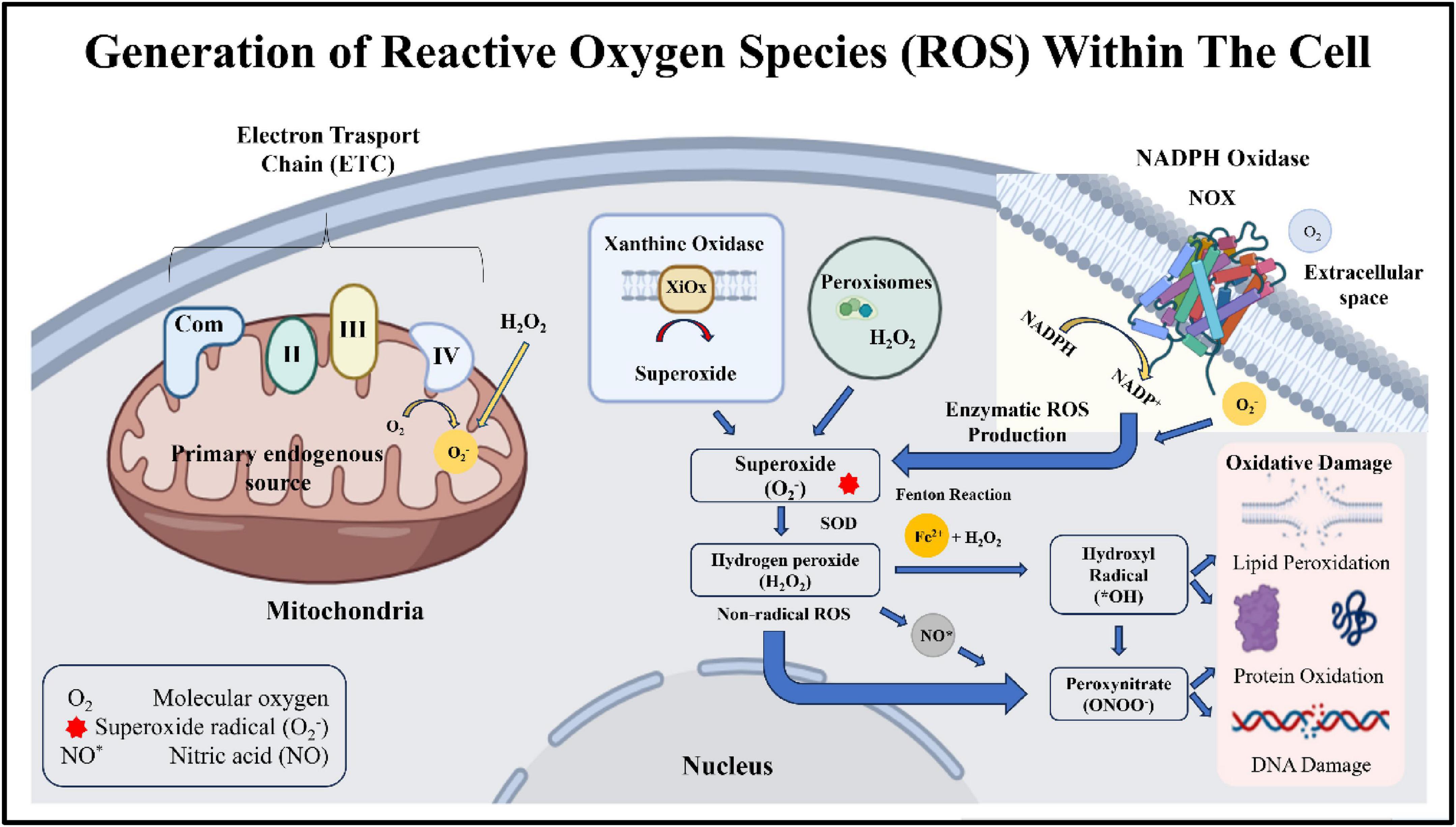
Figure 1. Generation of ROS within the cell.
Molecular damage induced by oxidative stress
High levels of ROS can cause severe damage to vital biomolecules, which in turn leads to biological dysfunction and the development of diseases (17). One of the major effects of oxidative stress is lipid peroxidation. During lipid peroxidation, ROS attack polyunsaturated fatty acids in the membranes of cells so as to generate lipid radicals and reactive aldehydes such as malondialdehyde (MDA) (23). As a result, the membrane loses its integrity, fluidity, and selective permeability. Besides lipids, protein molecules also undergo oxidative changes that can be detrimental. This results in their change of conformation, the disappearance of their enzymatic activities, and their becoming more prone to being broken down (24). Oxidative stress can cause proteins to become carbonylated, form disulfide bonds, and generate advanced oxidation protein products (24). Besides that, nucleic acids are susceptible to damage by ROS, resulting in changes of bases, breakage of strands, and mutations. For example, 8-hydroxy-2’-deoxyguanosine (8-OHdG), which is a result of guanine oxidation, is the widely known indicator of deoxyribonucleic acid (DNA) damage that is induced by ROS (Figure 2) (25). Altogether, such molecular changes are responsible for the aging of cells, programmed cell death, and the onset of several pathological conditions.
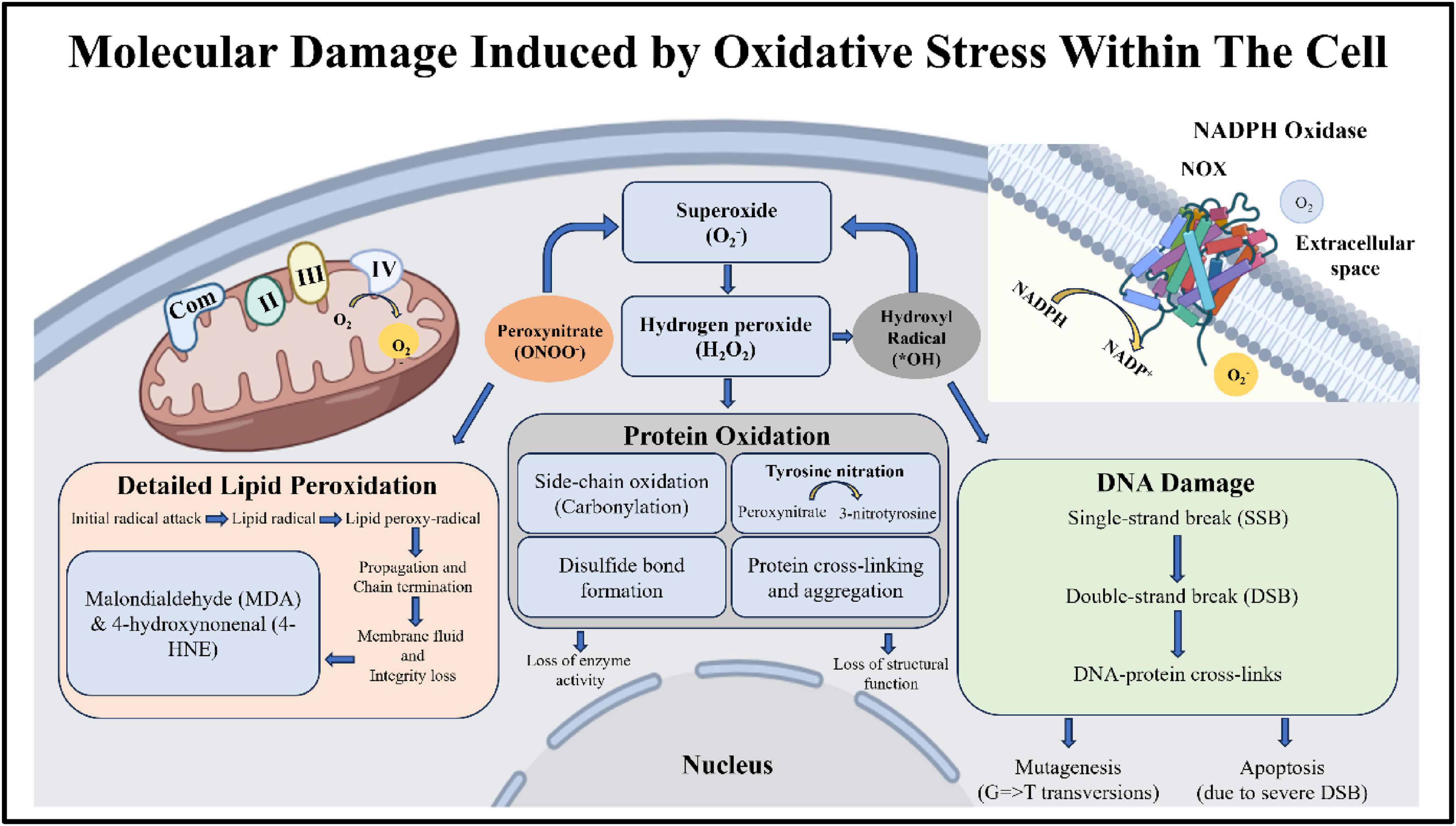
Figure 2. Molecular damage induced by oxidative stress.
Endogenous antioxidant defense systems
To compensate for the damaging effects of ROS, changes in organisms’ lives are possible. One of the changes is the evolution of a complex antioxidant defense system, which comprises two parts: enzymatic and non-enzymatic sectors (26). These protective mechanisms work in concord to maintain the redox balance and protect the cells from oxidative injury.
Enzymatic antioxidants
Enzymatic antioxidants primarily serve as a defense system against ROS. SOD is an enzyme that catalyzes the transformation of O2– into H2O2 and oxygen (27). This mechanism prevents the accumulation of superoxide radicals. Subsequently, catalase and GPx dispose of H2O2 by converting it into water and oxygen, thereby controlling the production of the highly reactive hydroxyl radical (28). Catalase occurs predominantly in peroxisomes and is highly efficient in decomposing H2O2 (29). Contrarily, GPx, in association with reduced GSH, detoxifies H2O2 and lipid hydroperoxides (Figure 3). GSH reductase ensures continuous availability of GSH, which is a very critical antioxidant by converting glutathione disulfide (GSSG) to GSH (30).
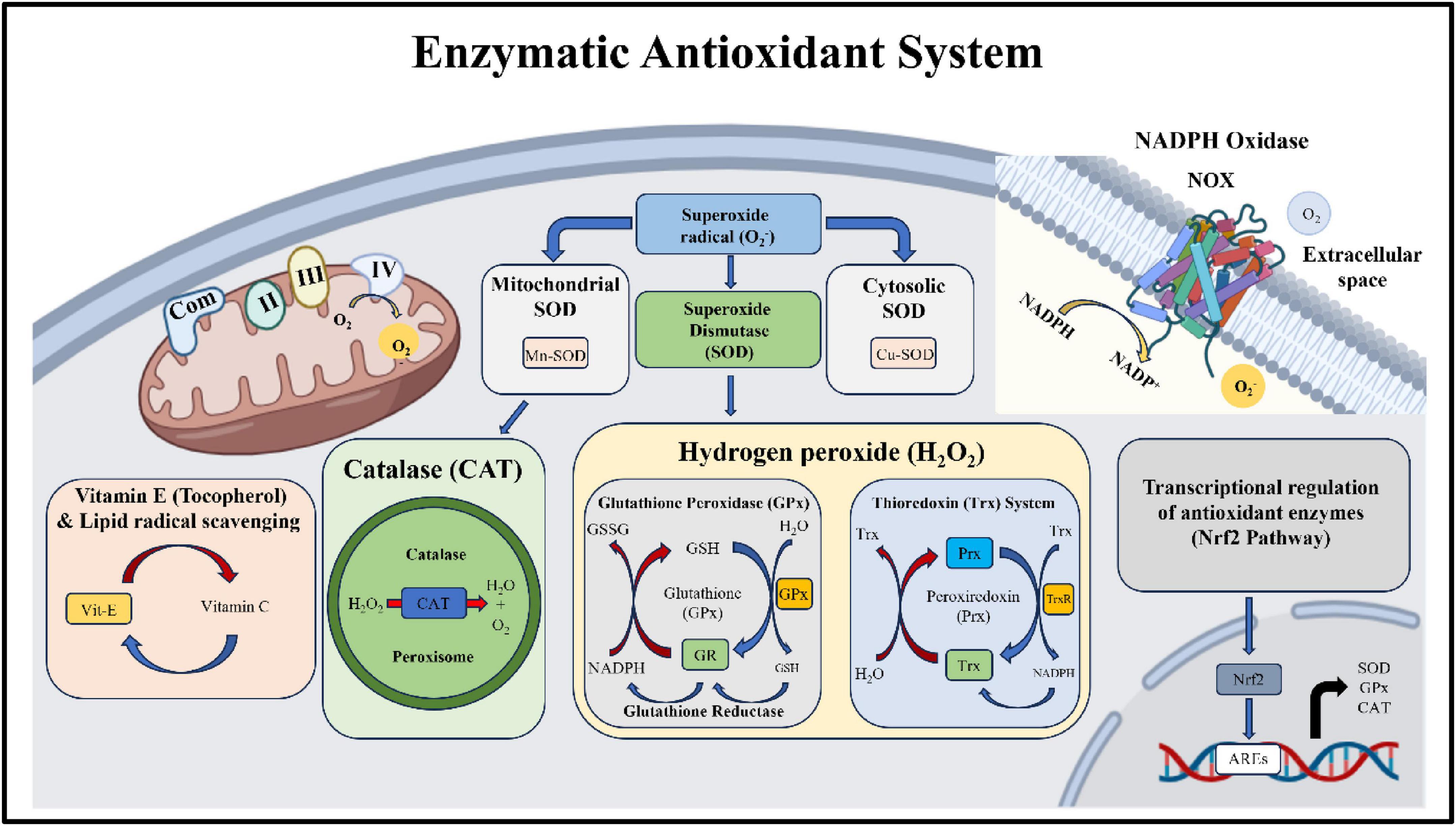
Figure 3. Enzymatic antioxidant system.
Non-enzymatic antioxidants
Non-enzymatic antioxidants complement enzymatic antioxidant systems by direct interaction with free radicals to neutralize them and terminate running chain reactions (31). They consist of the body’s own molecules such as GSH, uric acid, and coenzyme Q, as well as dietary antioxidants like vitamins C and E, carotenoids, and polyphenols (31). Vitamin C (ascorbic acid) is a very potent water-soluble antioxidant capable of dealing with a broad spectrum of ROS. At the same time, vitamin E (α-tocopherol) is the lipid-soluble antioxidant that safeguards the integrity of the membrane lipids by hampering the process of lipid peroxidation. Due to their electron donation and metal ion chelation abilities, polyphenolic compounds like flavonoids and phenolic acids have excellent antioxidant capabilities. Aside from neutralizing ROS, these compounds are also capable of influencing the cellular signaling pathways related to the response to oxidative stress.
Redox signaling and cellular adaptation
Besides causing damage, ROS may also act as signaling molecules that control various cellular activities. Redox-sensitive signaling pathways, including the Nrf2/Keap1 pathway, are key players in preserving cellular redox homeostasis (32). When exposed to oxidative stress, Nrf2 is released from Keap1 and moves into the nucleus, where it turns on the genes driven by antioxidant response element (ARE) (33). These genes produce a range of cytoprotective proteins such as antioxidant enzymes and phase II detoxification enzymes. Nuclear factor-kappa B (NF-κB), mitogen-activated protein kinases (MAPKs), and phosphoinositide 3-kinase/Akt (PI3K/Akt) are some of the other signaling pathways regulated by ROS (34). While low to moderate levels of ROS could induce beneficial cellular responses, high levels of ROS could disrupt these pathways, resulting in inflammation, apoptosis, and the progression of diseases (Figure 4).
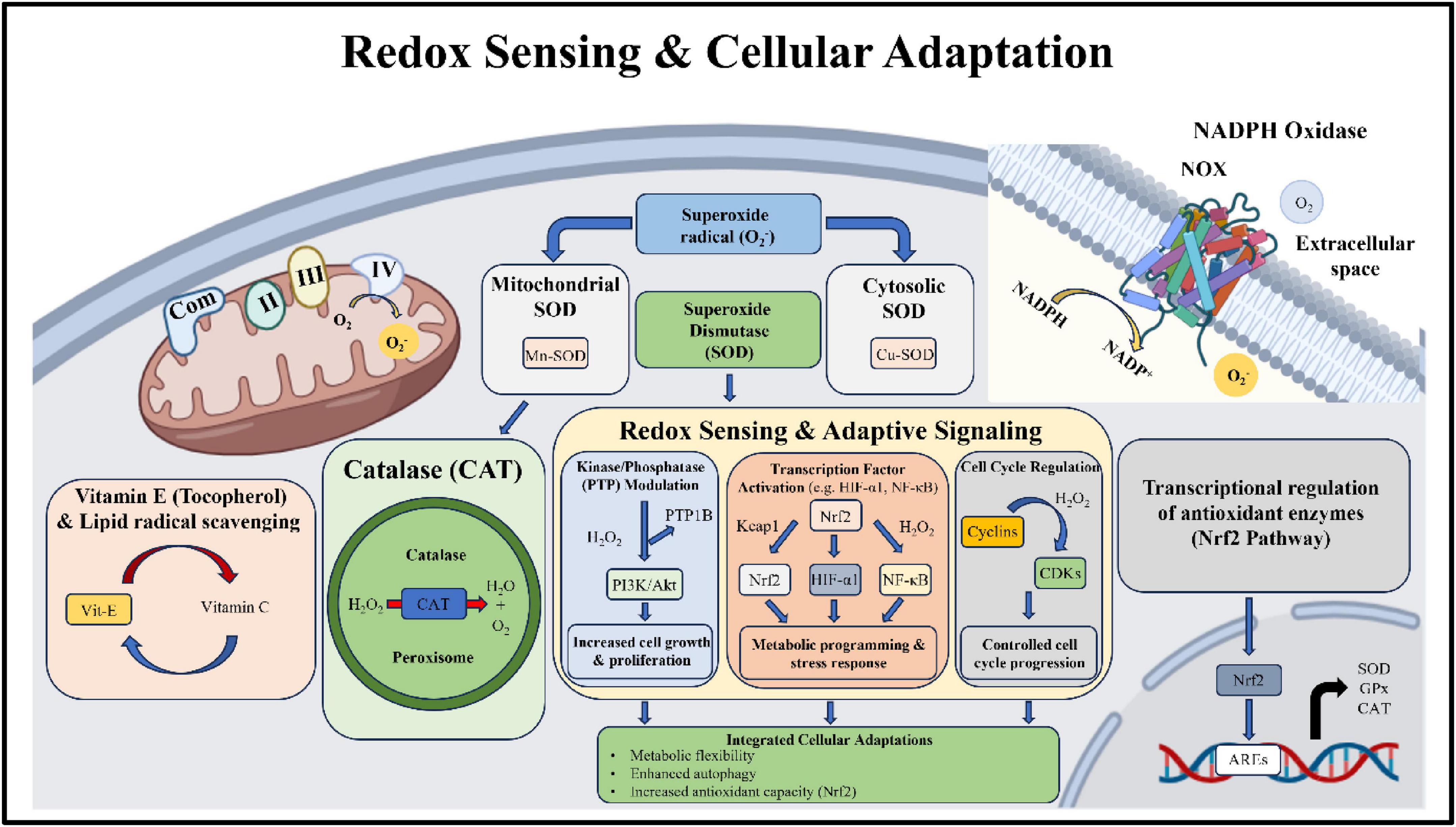
Figure 4. Redox sensing and cellular adaptation within the cell.
Imbalance and pathological implications
Oxidative stress occurs when the equilibrium of ROS generation and antioxidant defenses is disturbed, causing disease to result. Long-term oxidative stress is a great cause for major neurodegeneration, cardiovascular, diabetes, cancer, and aging-related complications (35). Excess ROS over a prolonged period of time will not only lead to impairment of the cellular structures, but a cascade of inflammatory reactions will be one of the consequences, leading to a cycle of oxidative damage that is very difficult to stop. Since endogenous defense systems are limited in their capability to control the oxidative stress and preserve redox homeostasis, the exploitation of therapeutic strategies is the only option to the greatest extent (36). As a result, a great number of researchers are currently focusing on the antioxidative systems based on nanoparticles, which are capable of more effective and selective intervention to oxidative stress.
Role of nanoparticles in antioxidant therapy
Nanotechnology is becoming a very effective method for overcoming the inherent problems of traditional antioxidant therapies (37). For instance, it allows you to have a very detailed control over the delivery, stability, and function of the therapeutic substances. Nanotechnology, a field employing particles that are usually 1–100 nm in size, offers unique physical and chemical properties like a large surface area relative to volume, a surface chemistry that can be modified quite easily, and an increased reactivity. These features make them very attractive for biomedical applications, particularly in fighting oxidative stress. With respect to antioxidant therapy, nanoparticles can play the role of carriers for antioxidant molecules, or they might also have the capability to directly get rid of the ROS themselves (38).
Types of nanoparticles used in antioxidant therapy
A wide range of nanoparticle systems has been explored for their antioxidant potential, each offering distinct advantages based on their composition, structure, and functional properties. Table 1 represents the application of nanoparticles in antioxidant therapy.
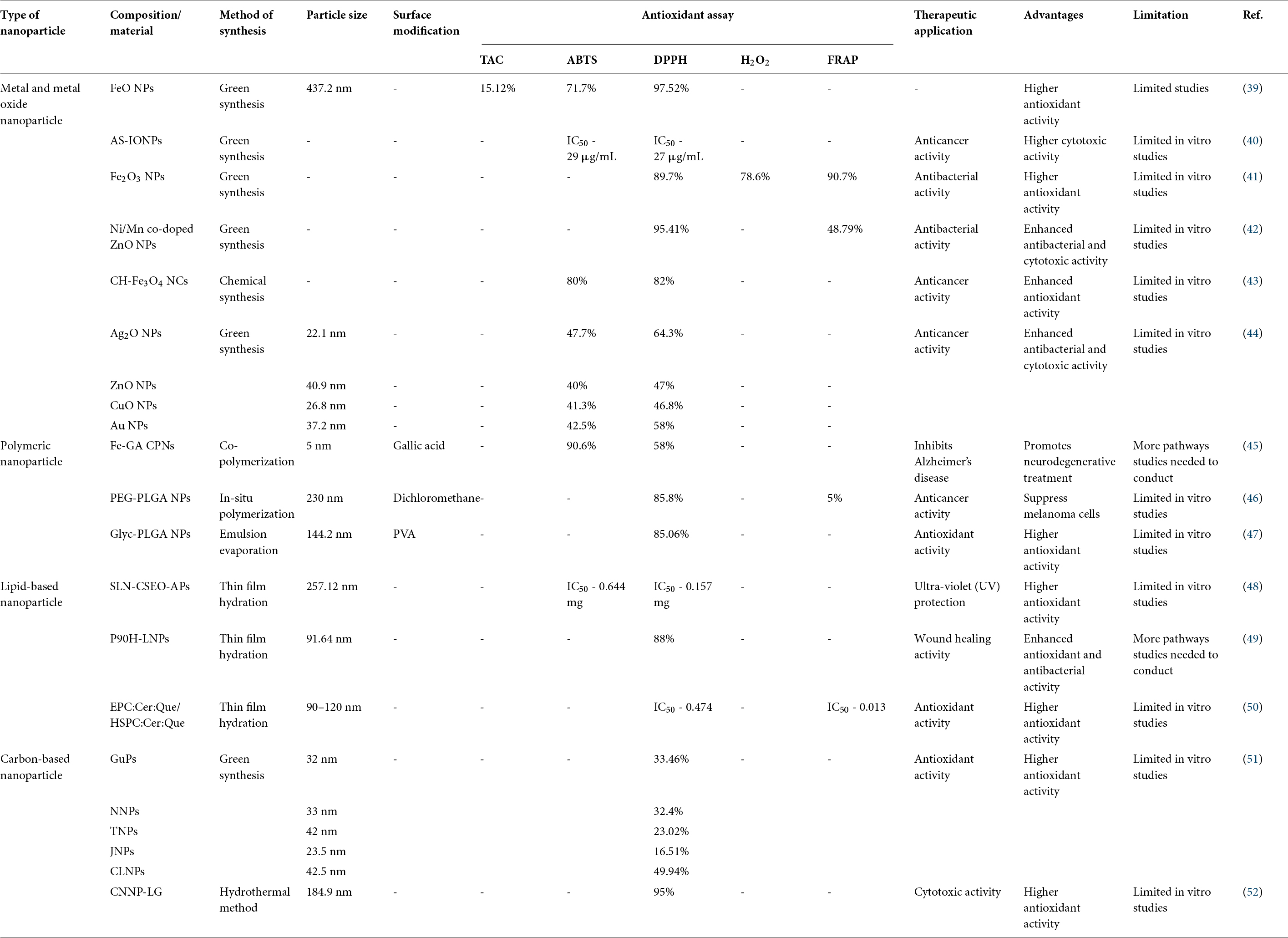
Table 1. Application of nanoparticles in antioxidant therapy.
Metal and metal oxide nanoparticles
Among all nanomaterials, metal oxide nanoparticles have been top choices in the antioxidant field for their redox-activated surfaces and enzyme-like properties. CeO2 nanoparticles, or nanoceria, are very special because of changing their oxidation states between Ce3+ and Ce4+ (10). This back and forth of redox cycling allows them to exhibit the functions of the major antioxidant enzymes, SOD and catalase, and thus help in effective ROS scavenging. Besides that, ZnO nanoparticles have been proved to have antioxidant and antiinflammatory effects, which, however, is often a matter of dosage (53). TiO2 nanoparticles, although frequently used in the biomedical field, need thorough surface coating so that there is no generation of pro-oxidant effects under UV light (54). These nanoparticles can be developed to show regulated antioxidant characteristics by means of doping and surface functionalization.
Polymeric nanoparticles
Polymeric nanoparticles are among the most popular delivery systems for antioxidant compounds, mainly due to their biocompatibility, biodegradability, and flexibility in terms of design (55). Polymers such as poly(lactic-co-glycolic acid) (PLGA), chitosan, polyvinylpyrrolidone (PVP), and hydroxypropyl cellulose (HPC) are the most suitable materials to deliver curcumin resveratrol quercetin, and other bioactive substances (56). One of the major advantages of encapsulation is that it provides a physical barrier against reacting with the external environment, thereby preventing breakage of antioxidants, better solubilization, and regulated and prolonged liberation of active agents. Moreover, polymeric nanoparticles can be equipped with the targeting ligands, which enable them to deliver drugs to the specific area or site in the body (57). This way, therapeutic effectiveness can be greatly enhanced, and at the same time, systemic side effects can be reduced to a minimum.
Lipid-based nanoparticles
Delivering both water-soluble and lipophilic antioxidants has been made possible by lipid-based nanoparticles such as liposomes, solid lipid nanoparticles (SLNs), and nanostructured lipid carriers (NLCs) (58). Besides being very biocompatible and having a low toxicity, these delivery systems are highly regarded from a clinical point of view. Liposomes that consist of phospholipid bilayers are very good at targeting antioxidant molecules to specific tissues, whereas SLNs and NLCs offer better storage stability and are able to release drugs in a controlled manner (59). Lipid-based nanoparticles are the most suitable method for enhancing the bioavailability of poorly soluble antioxidants and also help in transporting them through the biological barriers.
Carbon-based nanomaterials
Carbon-based nanomaterials like graphene oxide, reduced graphene oxide, fullerenes, and carbon nanotubes have been found to possess antioxidant properties due to their extraordinary electronic structures and enhanced ability to react with their environment at the surface level (60). By means of electron transfer, these materials are capable of neutralizing free radicals and serve as radical sponges. Fullerenes are capable of eradicating multiple ROS and that makes them “radical scavengers” as they display an unparalleled level of hydrogen capacity. However, taking into consideration the biocompatibility and the potential for long-term toxicity of fullerenes, this means that they may have a negative impact on the human body to some extent (61). As a result, safe biomedical application requires the performance of surface modification and functionalization along with other measures to achieve this goal.
Synthesis strategies for antioxidant nanoparticles
The synthesis is done really influences the physicochemical characteristics, stability, and even the bio-activity of the nanoparticles. Many methods have been designed to create nanoparticles possessing specific antioxidant traits.
Chemical and physical methods
Classic methods for synthesis like solgel methods, hydrothermal ways of production, and chemical reduction provide accurate control over the particle’s size, shape, and composition (62). These techniques are the most common ways of making metal and metal oxide nanoparticles with specific features. On the other hand, they require the usage of toxic substances and very strong reaction conditions that may prevent the applicability of the materials for biomedical uses (63).
Green synthesis method
Green synthesis is a method that has become a very popular environmentally friendly and sustainable alternative for the fabrication of nanoparticles (64). Such a method endeavors biological entities such as plant extracts, microorganisms, and biomolecules while acting as reducing and stabilizing agents. The phytochemicals, such as flavonoids, phenolics, and alkaloids, are the main factors that help the reduction of metal ions and cap the resulting nanoparticles (65). Nanoparticles that are green-synthesized frequently have better biocompatibility and inherent antioxidant properties, which is because of the existence of the bioactive compounds on the surface of the nanoparticles. This approach is compatible with the principles of green chemistry and is playing an ever more important role in biomedical research.
Mechanisms of nanoparticle- mediated antioxidant activity
Besides regular antioxidants, nanoparticle antioxidant therapy uses a whole range of mechanisms that are based on the peculiar physicochemical properties of nanoparticles, such as extremely high surface reactivity, tunable redox potential, and capability to interact with biological molecules (66). Typically, antioxidants of nanoparticles can be divided into four main roles: direct neutralization of ROS, catalysis by nanozymes, localized delivery of antioxidant molecules, and alteration of cellular signaling pathways that depend on redox state.
Direct ROS scavenging
One of the major mechanisms by which nanoparticles exert their antioxidant effects is the scavenging of ROS directly. In fact, the very high surface areas coupled with highly reactive surfaces of the nanoparticles make it possible for them to transfer electrons or hydrogen atoms to free radicals, which leads to their stabilization and cessation of the propagation of oxidative damage chain reactions (38). This is particularly crucial in the prevention of lipid peroxidation and ensuring the stability of cellular membranes. Actually, metal and carbon-based nanoparticles are very efficient in directly cleaving out ROS (67). Metal oxide nanoparticles create surface defects and oxygen vacancies that enhance their interaction with ROS, and also, they participate in electron transfer reactions that transform reactive species to less harmful ones (68). In a like manner, carbon-based nanomaterials, including fullerenes and graphene derivatives, have been given the title of “radical sponges” since they can adsorb and neutralize several ROS molecules at the same time (69). This capability of the nanoparticles for the indiscriminate scavenging phenomenon sets them apart from natural antioxidants, which usually target specific types of ROS.
Enzyme-mimetic activity
One of the remarkable features of some nanoparticles is their ability to carry out the catalytic roles of natural antioxidant enzymes, which has led to their being identified as “nanozymes.” Through this process, nanoparticles catalytically degrade ROS just like our body’s own defense systems; thus, they can provide ongoing and highly effective antioxidant protection (70). This back-and-forth shift enables them to adequately perform SOD-like functions by changing superoxide radicals into H2O2 and also play a catalase-like role by converting H2O2 to water and oxygen (Figure 5) (29). Notably, these nanoparticles are regenerable, which means that they can undergo a series of redox reactions over and over without being depleted.
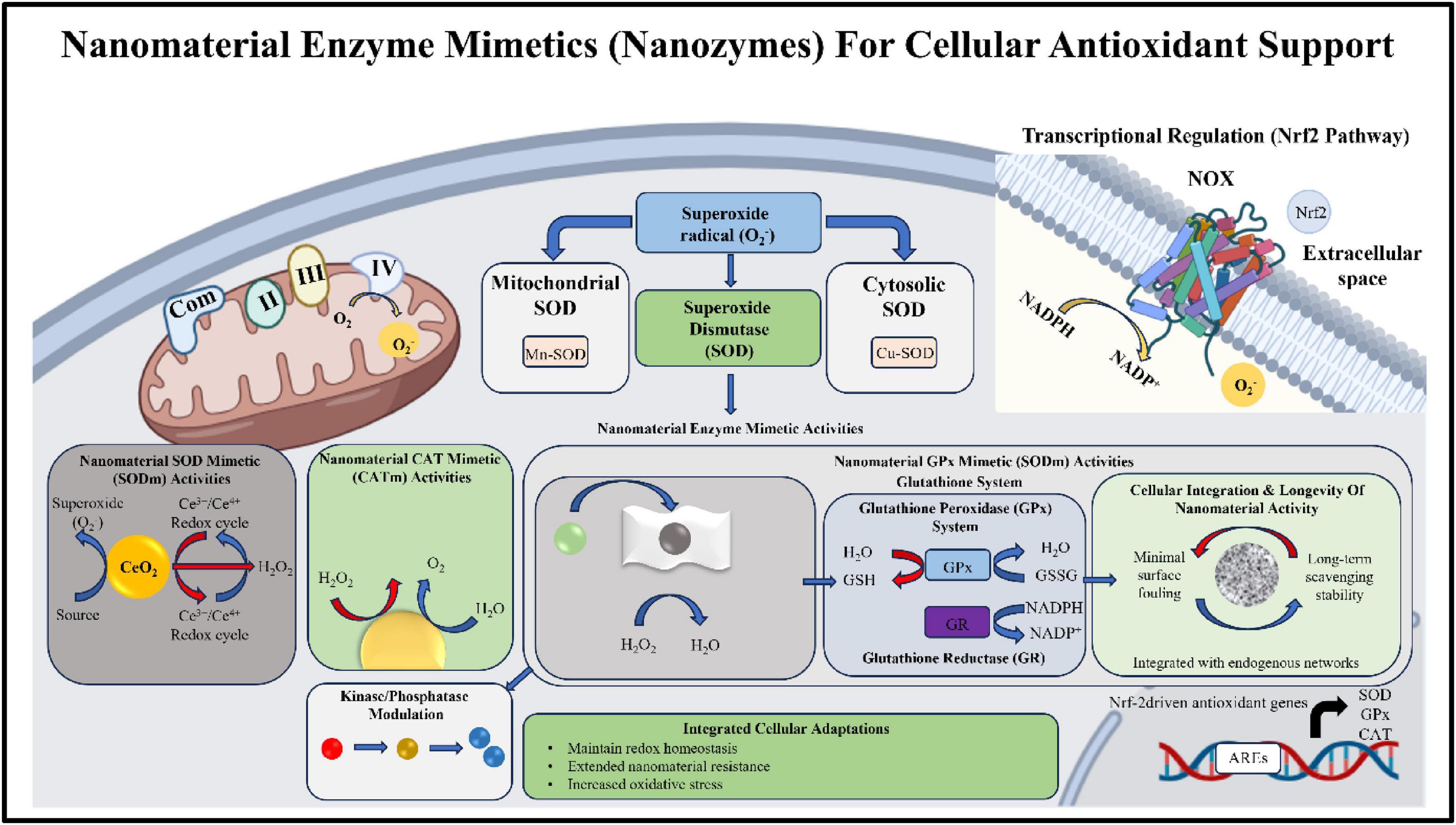
Figure 5. Nanomaterial enzyme mimetics for cellular antioxidant support.
Controlled delivery of antioxidant agents
Besides being highly bioactive on their own, nanoparticles are excellent carriers for transporting various antioxidant molecules right where they are needed in the body. Many traditional antioxidants like curcumin, resveratrol, and quercetin have issues like low water solubility, fast breakdown and low absorption in the body (71). Encapsulation in nanoparticles is one of the strategies to address these problems, as the carriers can protect the active substances from destruction too early and also enhance the pharmacokinetic properties of such compounds (72). More significantly, nanoparticle delivery systems can offer a controlled release of antioxidants in a continuous manner, which not only maintains the therapeutic level of antioxidants for a longer time but also lowers the frequency of drug administration (73). Additionally, nanoparticle surfaces can be “decorated” with targeting ligands like antibodies, peptides, or small molecules. Such an “artificial” surface enables site-specific delivery at main oxidative stress sites, e.g., inflamed tissues or tumor microenvironments. Table 2 emphasizes the controlled delivery of antioxidant agents using nanotechnology-based delivery systems.
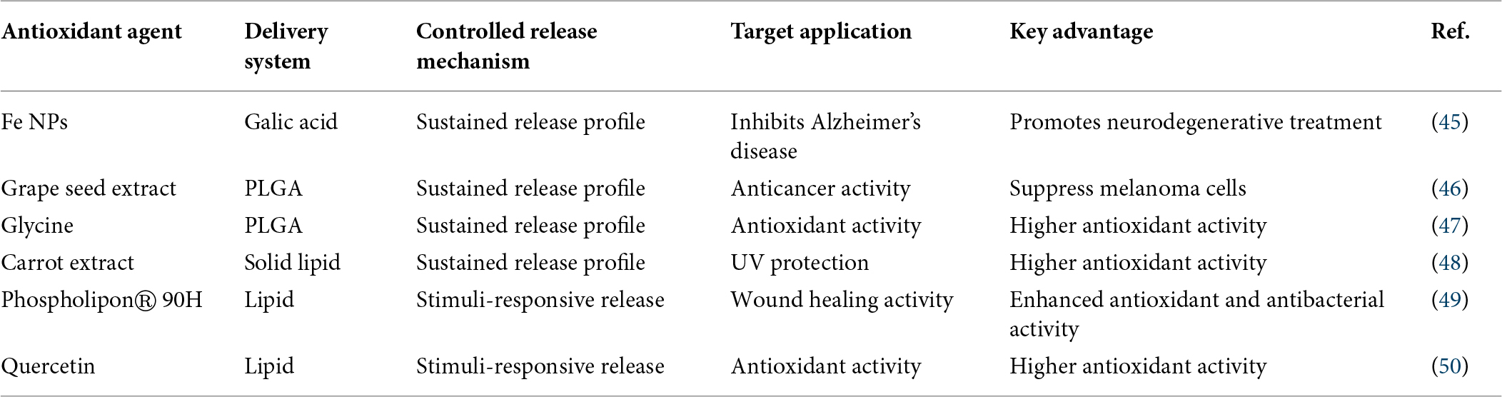
Table 2. Controlled delivery of antioxidant agents using nanotechnology-based delivery systems.
Modulation of redox-sensitive cellular signaling pathways
Apart from the direct ROS neutralization, nanoparticles also have the ability to alter intracellular signaling pathways that regulate oxidative stress responses. In this context, the Nrf2/Keap1 pathway is one of the most crucial pathways. Activated Nrf2 induces the transcription of a wide range of antioxidant and cell-protective genes, e.g., those encoding SOD, catalase, GPx, and phase II detoxification enzymes (74). It is known that some nanoparticles contribute to the release of Nrf2 from Keap1, its cytoplasmic inhibitor, which then allows its nuclear translocation and activation of AREs (75). This mechanism of action not only remarkably enhances the cells’ intrinsic antioxidant capacity but also prepares them to endure oxidative damage for the longer term. In addition to the Nrf2 pathway, nanoparticles can also influence other redox-sensitive signaling pathways, like NF-B, MAPKs, and PI3K/Akt (76). Nanoparticles, by modulating these pathways, can lead to the suppression of inflammation, decrease of apoptosis, and promotion of cell survival and tissue regeneration.
Regeneration of endogenous antioxidant systems
Besides, nanoparticles contribute significantly to triggering the body’s natural antioxidant defenses and restoring them. Certain nanoparticle varieties can raise intracellular GSH levels, which is a crucial non-enzymatic antioxidant (28). They may achieve this by either promoting its synthesis or by transforming its oxidized form GSSG back to GSH. The primary results of this procedure include sustaining cellular redox homeostasis in combination with increased antioxidant capacity. Additionally, nanoparticles could serve as a vehicle to protect antioxidant enzymes from their oxidative modification and thereby preserve their enzymatic activity (77). The synergistic interaction of nanoparticle-based treatments with endogenous defense mechanisms results in more powerful overall therapeutic effects.
Biomedical applications
The special feature of nanoparticles to influence oxidative stress in more than one way has made them very popular and versatile in different biomedical areas. Besides having natural antioxidant effects to fight free radicals, nanoparticles can also deliver drugs to specific sites and release them in a controlled manner, which makes them very useful for the treatment of several diseases caused by oxidative stress. An overview of the major biomedical uses of antioxidant therapy via nanoparticles, with emphasis on the main diseases where oxidative stress is an important factor in the pathogenesis.
Wound healing
Oxidative stress can be a major barrier to wound healing, as it fosters inflammation, hampers cell growth, and stops new tissue formation. Too many ROS at the site of the wound pose a risk to cellular elements and can break down the extracellular matrix, which will lead to an extended inflammatory period and postponed healing (78). Metal oxide nanoparticles such as cerium and zinc oxides have the ability to scavenge ROS, and along with them, they have antiinflammatory and antimicrobial properties that contribute to creating a healing-friendly microenvironment (79). Alongside this, nanoparticle-loaded polymeric scaffolds and nanofibers not only offer a framework but also enable the therapeutic agents to be released in a controlled manner (9, 80). These can promote new blood vessel formation, increase fibroblast cell production, and hasten the top layer of skin coming together.
Cancer therapy
Oxidative stress in cancer is a double-edged sword, as it helps in tumor progression as well as therapeutic response. Tumors can proliferate and survive due to moderate ROS levels, whereas extra ROS can cause cancer cells’ death (81). Antioxidant therapy catalyzed by nanoparticles presents a unique way of finely tuning ROS levels. Nanoparticles have the potential to be designed to impart antioxidant drugs to tumors only, consequently sparing healthy cells from oxidative damages occurring during chemotherapy and radiotherapy (76). At the same time, some nanoparticle systems can raise oxidative stress within only cancer cells, killing them by apoptosis and increasing therapeutic effects. Such dual functions thus allow a balanced way of handling cancer treatment.
Neurodegenerative diseases
Oxidative stress-induced neuronal damage has been identified as a main cause for neurodegenerative disorders such as Alzheimer’s disease, Parkinson’s disease, and Huntington’s disease (82). The brain is more susceptible to oxidative stress since it uses a lot of oxygen, has the highest quantity of lipids, and has a comparatively low level of antioxidants. Nanoparticles provide a very promising way for neuroprotection since they can efficiently scavenge ROS and also deliver drugs across the blood-brain barrier (BBB) (83). Lipid-based and polymeric nanoparticles could be modified in such a way so that they cross the BBB and simultaneously release antioxidants within the central nervous system (37). One of the main reasons why cerium oxide nanoparticles exert such neuroprotective effects is their ability to constantly regenerate antioxidant activity (84). These nanoparticles not only decrease neuronal oxidative damage, but they also suppress inflammation and enhance mitochondrial function. Therefore, nanoparticle-facilitated antioxidant therapy is a very good candidate for slowing down disease spreading and enhancing neurological function.
Cardiovascular diseases
Oxidative stress plays a significant role in the onset of cardiovascular diseases such as atherosclerosis, hypertension, and myocardial infarction (85). One of the major pathological processes caused by ROS in these diseases is vascular endothelial dysfunction, the oxidation of lipids, and turbulent inflammatory responses (86). Nanoparticles provide a potential strategy for the treatment of oxidative stress in the cardiovascular system. Besides their ability to scavenge ROS, they are also capable of delivering antioxidants to the specific sites of injury. Actually, nanoparticle-based drug delivery systems have the ability to target atherosclerotic plaques directly, in addition to helping repair oxidative and inflammatory injuries of the vascular walls (87). This form of targeted delivery can make a treatment more effective while at the same time reducing the exposure of the rest of the body to the drug. Nanoparticles are not only going to reduce oxidative stress. They can help cardiovascular drugs to work better by not only keeping them safe from being broken down but also by helping them get into the bloodstream. Besides this, their ability to influence the redox-sensitive signaling pathways is the main reason for their continual beneficial effects on cardiovascular health.
Diabetes and metabolic disorders
Chronic oxidative stress is a major factor in the development of diabetes and its related complications, including diabetic neuropathy, nephropathy, and retinopathy (88). Excessive ROS production induced by hyperglycemia disrupts cells, causes inflammation, and affects the functioning of tissues (89). A new and promising method to reduce oxidative stress in diabetes is the use of nanoparticles as a carrier system for antioxidant drugs. With these nanoparticles, antioxidants can be precisely delivered to the diabetes-affected tissues that include the islets of Langerhans, kidneys, and peripheral nerves (90). Targeted delivery of antioxidants leads not only to the dramatic reduction of tissue damage but also assists in the recovery of cellular functions. Moreover, nanomaterials for drug delivery systems in diabetic wound healing have remarkable potential in enhancing tissue regeneration and infection control. Furthermore, by altering the redox signaling pathways, nanoparticles can act not only to regulate glucose metabolism and insulin sensitivity but also to aid in metabolic homeostasis.
Toxicological considerations and limitations
Antioxidant systems based on nanoparticles have the potential to be very effective therapeutic agents. However, these treatment agents have not reached clinical use to date mainly because certain drawbacks and toxicological issues need to be deeply understood and resolved clearly. It is reported that the biological effect of nanoparticles mostly depends on their natural physicochemical features such as size, shape, surface charge, composition, and concentration. Indeed, these characteristics not only control the ability of particles to be taken up by cells and their localization within the organism but also their interaction with biological components, all of which together define their safety profile. Besides safety, one of the major concerns of nanoparticle use is the induction of toxicity, which could be the consequence of the particles stealthily interacting with cell components. For instance, because of their greater surface reactivity, small nanoparticles can penetrate the cell membranes and accumulate in organs such as the liver, kidneys, and brain. Even more, under certain conditions, nanoparticles may behave as pro-oxidants leading to ROS production instead of their removal, which would result in elevated oxidative stress and cell damage. Another big concern is a lack of adequate elimination of nanoparticles. If nanoparticles do not break down sufficiently and stay present within the tissues, they might lead to persistent toxicity and inflammation. On the other hand, proteins that are adsorbed, forming a corona around the nanoparticles in biological fluids, may alter their surface properties, which could reduce the efficiency in targeting cells and might also trigger an immune response.
Future perspectives
Currently, therapies based on nanoparticles for the delivery of antioxidants are evolving rapidly and have huge potential to completely change the way we treat diseases that involve oxidative stress. It’s very likely that in the next few years research will focus on the development of smart nanoparticles capable of responding to biological signals, e.g., by releasing drugs triggered by changes in pH, redox gradients, or enzymatic activity. This type of mechanism would provide a very high degree of drug release control in terms of both time and location, resulting not only in a more potent therapeutic effect but also in the reduction of side effects. Additionally, the combination of diagnostic and therapeutic nanoparticles into a single system also seems very promising. This dual-function approach could make it possible to determine the degree of oxidative stress while at the same time delivering the medication directly to the source. Also, developments in surface modifications and ligand alterations can enhance targeting features, which help nanoparticles to home in on specific disease locations. At the same time, a rise in personalized nanomedicine applications, where nanoparticle formulas are personalized based on patient-specific parameters, disease characteristics, and genomics. Here, a personalized therapy approach not only has the capability to improve efficacy significantly but also to reduce differences in patient responses. Besides that, environmentally friendly production and the use of biocompatible materials are likely to focus safety aspects and the sustainability of nanoparticle manufacturing. Actual clinical trials will have to address issues such as scale-up production, regulatory approval, and extensive toxicological testing, among others.
Conclusion
Nanoparticle-based antioxidant therapy is a very suitable and highly promising method, especially since oxidative stress is still considered the primary factor responsible for many chronic and degenerative diseases. Through the use of the unique physicochemical properties of nanoparticles, these platforms are able to overcome several limitations of conventional antioxidants such as their low efficiency, instability, and lack of specificity. In fact, the ability of nanoparticles to combine different approaches such as direct ROS scavenging, nanozyme activity, and drug release, as well as modulating redox-sensitive pathways, makes them the future of therapeutic tools in the domain of biomedicine. Biomedical applications, such as wound healing, cancer treatment, and neurodegenerative and heart diseases, are a testament to how nanoparticle-based antioxidant systems have become an essential part of the clinics. Not only has their capability to boost the therapeutic effect been proven, but their multifunctional nature has also endowed them with the power to carry out targeted and long-term therapy, which is indeed quite necessary when one has to deal with very glycemic diseases. However, besides these fundamental breakthroughs, the problems of toxicity, safety over the long term, body distribution, and the obtaining of regulations are still the main factors limiting the extensive use of these therapies in the clinical setting. These defects in large part stand to be removed by the use of standardized production procedures, targeted toxicological studies, and the development of materials that are both compatible and degradable. Eventually, nanoparticle-based therapies for antioxidants present a radical method that can change the handling of oxidative stress-related diseases. Only cross-disciplinary research and technological breakthroughs will pave the way for these promising systems to be turned from experimental models to safe and effective clinical applications.
Ethics statement
Not applicable.
Funding
The authors declare that no funds, grants, or other support were received during the preparation of this manuscript.
Conflict of interest
The authors declare that the research was conducted in the absence of any commercial or financial relationships that could be construed as a potential conflict of interest.
References
1. Fathi A, Shiade SRG, Saleem A, Shohani F, Fazeli A, Riaz A , et al. Reactive oxygen species (ROS) and antioxidant systems in enhancing plant resilience against abiotic stress. Int J Agron. (2025) 2025(1):8834883. doi: 10.1155/ioa/8834883
2. Zandi P, Schnug E. Reactive oxygen species, antioxidant responses and implications from a microbial modulation perspective. Biology (Basel). (2022) 11(2):155. doi: 10.3390/biology11020155
3. Andrés CMC, Pérez de la Lastra JM, Juan CA, Plou FJ, Pérez-Lebeña E. The role of reactive species on innate immunity. Vaccines (Basel). (2022) 10(10):1735. doi: 10.3390/vaccines10101735
4. Perluigi M, Di Domenico F, Butterfield DA. Oxidative damage in neurodegeneration: roles in the pathogenesis and progression of Alzheimer disease. Physiol Rev. (2024) 104(1):103–97. doi: 10.1152/physrev.00030.2022
5. Engwa GA, Nweke FN, Nkeh-Chungag BN. Free radicals, oxidative stress-related diseases and antioxidant supplementation. Alternat Ther Health Med. (2022) 28(1):114–28.
6. Liu Y, Wu G, Feng L, Li J, Xia Y, Guo W , et al. Harnessing antioxidants in cancer therapy: opportunities, challenges, and future directions. Antioxidants (Basel). (2025) 14(6):674. doi: 10.3390/antiox14060674
7. Szczyglewska P, Feliczak-Guzik A, Nowak I. Nanotechnology-general aspects: a chemical reduction approach to the synthesis of nanoparticles. Molecules. (2023) 28(13):4932. doi: 10.3390/molecules28134932
8. Stevanović M, Filipović N. A review of recent developments in biopolymer nano-based drug delivery systems with antioxidative properties: insights into the last five years. Pharmaceutics. (2024) 16(5):670. doi: 10.3390/pharmaceutics16050670
9. Selvaraj S, Subbarayan R, Chauhan A, Jabir M, Ghotekar S. MXene-loaded hydrogels for tissue regeneration: a review of recent advances and future perspectives. J Mater Chem B. (2026) 14:3972–93. doi: 10.1039/d5tb02696b
10. Selvaraj S, Chauhan A, Radhakrishnan A, Rana G, Dutta V, Batoo KM , et al. Cerium oxide nanoparticles and their polymeric composites: advancements in biomedical applications. J Inorgan Organomet Poly Mater. (2024) 34(12):5691–717. doi: 10.1007/s10904-024-03263-5
11. Selvaraj S, Dutta V, Gopalakrishnan C, Subbarayan R, Rana G, Radhakrishnan A , et al. Biomedical potential of hydrogels: a multifaceted approach to innovative medication delivery. Emerg Mater. (2024) 7(3):721–63. doi: 10.1007/s42247-024-00643-y
12. Pandey V, Pandey T. A mechanistic understanding of reactive oxygen species (ROS)-responsive bio-polymeric nanoparticles: current state, challenges and future toward precision therapeutics. Biopolymers. (2025) 116(3):e70027. doi: 10.1002/bip.70027
13. Mukherjee AG, Gopalakrishnan AV. The mechanistic insights of the antioxidant Keap1-Nrf2 pathway in oncogenesis: a deadly scenario. Med Oncol. (2023) 40(9):248. doi: 10.1007/s12032-023-02124-4
14. Wu J, Zhu Z, Liu W, Zhang Y, Kang Y, Liu J , et al. How nanoparticles open the paracellular route of biological barriers: mechanisms, applications, and prospects. ACS Nano. (2022) 16(10):15627–52. doi: 10.1021/acsnano.2c05317
15. Khan IA, Yu T, Yang M, Liu J, Chen ZA. Systematic review of toxicity, biodistribution, and biosafety in upconversion nanomaterials: critical insights into toxicity mitigation strategies and future directions for safe applications. BME Front. (2025) 6:0120. doi: 10.34133/bmef.0120
16. Abram SL, Tavernaro I, Johnston LJ, Zou S, Resch-Genger U. Nanoscale reference and test materials for the validation of characterization methods for engineered nanomaterials - current state, limitations, and needs. Anal Bioanal Chem. (2025) 417(12):2405–25. doi: 10.1007/s00216-024-05719-6
17. Jomova K, Raptova R, Alomar SY, Alwasel SH, Nepovimova E, Kuca K , et al. Reactive oxygen species, toxicity, oxidative stress, and antioxidants: chronic diseases and aging. Arch Toxicol. (2023) 97(10):2499–574. doi: 10.1007/s00204-023-03562-9
18. Deshpande OA, Mohiuddin SS. Biochemistry, oxidative phosphorylation. In: StatPearls. StatPearls Publishing (2023).
19. Letts JA, Sazanov LA. Clarifying the supercomplex: the higher-order organization of the mitochondrial electron transport chain. Nat Struct Mol Biol. (2017) 24(10):800–8. doi: 10.1038/nsmb.3460
20. Hanukoglu I. Antioxidant protective mechanisms against reactive oxygen species (ROS) generated by mitochondrial P450 systems in steroidogenic cells. Drug Metab Rev. (2006) 38(1–2):171–96. doi: 10.1080/03602530600570040
21. Fujii J, Homma T, Osaki T. Superoxide radicals in the execution of cell death. Antioxidants (Basel). (2022) 11(3):501. doi: 10.3390/antiox11030501
22. Kehrer JP. The Haber-Weiss reaction and mechanisms of toxicity. Toxicology. (2000) 149(1):43–50. doi: 10.1016/s0300-483x(00)00231-6
23. Onur Yaman S, Ayhanci A. Lipid peroxidation. In: Atukeren P editor. Accenting Lipid Peroxidation. London: IntechOpen (2021). p. 1–11.
24. Gęgotek A, Skrzydlewska E. Biological effect of protein modifications by lipid peroxidation products. Chem Phys Lipids. (2019) 221:46–52. doi: 10.1016/j.chemphyslip.2019.03.011
25. Goriuc A, Cojocaru KA, Luchian I, Ursu RG, Butnaru O, Foia L. Using 8-hydroxy-2’-deoxiguanosine (8-OHdG) as a reliable biomarker for assessing periodontal disease associated with diabetes. Int J Mol Sci. (2024) 25(3):1425. doi: 10.3390/ijms25031425
26. Bandodkar VV, Moger VS, Baliga P. Antioxidant defense mechanisms: enzymatic and non-enzymatic. In: Prabhakar PK editor. The Role of Reactive Oxygen Species in Human Health and Disease. IGI Global Scientific Publishing (2025). p. 43–80.
27. Anwar S, Sarwar T, Khan AA, Rahmani AH. Therapeutic applications and mechanisms of superoxide dismutase (SOD) in different pathogenesis. Biomolecules. (2025) 15(8):1130. doi: 10.3390/biom15081130
28. Jomova K, Alomar SY, Alwasel SH, Nepovimova E, Kuca K, Valko M. Several lines of antioxidant defense against oxidative stress: antioxidant enzymes, nanomaterials with multiple enzyme-mimicking activities, and low-molecular-weight antioxidants. Arch Toxicol. (2024) 98(5):1323–67. doi: 10.1007/s00204-024-03696-4
29. Xu D, Wu L, Yao H, Zhao L. Catalase-like nanozymes: classification, catalytic mechanisms, and their applications. Small. (2022) 18(37):e2203400. doi: 10.1002/smll.202203400
30. Averill-Bates DA. The antioxidant glutathione. Vitam Horm. (2023) 121:109–41. doi: 10.1016/bs.vh.2022.09.002
31. de Oliveira JM, Carnib BL, Destro ALF, Floresta LRS, Lage TM, dos Santos RM , et al. Non-enzymatic antioxidants and the regulation of redox balance in health: mechanisms and therapeutic implications. In: Reactive Oxygen Species – An Overview. London: IntechOpen (2026).
32. Verza FA, Da Silva GC, Nishimura FG. The impact of oxidative stress and the NRF2-KEAP1-ARE signaling pathway on anticancer drug resistance. Oncol Res. (2025) 33(8):1819–34. doi: 10.32604/or.2025.065755
33. Bian DD, Zhang X, Zhu XR, Tang WH, Peng Q, Chen YH , et al. The Nrf2-Keap1/ARE signaling pathway in aquatic animals. Int J Biol Macromol. (2025) 308:142595. doi: 10.1016/j.ijbiomac.2025.142595
34. Yang D, Yang L, Cai J, Li H, Xing Z, Hou Y. Phosphoinositide 3-kinase/Akt and its related signaling pathways in the regulation of tumor-associated macrophages polarization. Mol Cell Biochem. (2022) 477(10):2469–80. doi: 10.1007/s11010-022-04461-w
35. Yang J, Luo J, Tian X, Zhao Y, Li Y, Wu X. Progress in understanding oxidative stress, aging, and aging-related diseases. Antioxidants (Basel). (2024) 13(4):394. doi: 10.3390/antiox13040394
36. Forman HJ, Zhang H. Targeting oxidative stress in disease: promise and limitations of antioxidant therapy. Nat Rev Drug Discov. (2021) 20(9):689–709. doi: 10.1038/s41573-021-00233-1
37. Martinelli C, Pucci C, Battaglini M, Marino A, Ciofani G. Antioxidants and nanotechnology: promises and limits of potentially disruptive approaches in the treatment of central nervous system diseases. Adv Healthc Mater. (2020) 9(3):e1901589. doi: 10.1002/adhm.201901589
38. Samrot AV, Ram Singh SP, Deenadhayalan R, Rajesh VV, Padmanaban S, Radhakrishnan K. Nanoparticles, a double-edged sword with oxidant as well as antioxidant properties—a review. Oxygen. (2022) 2(4):591–604. doi: 10.3390/oxygen2040039
39. Nughwal A, Bharti R, Thakur A, Verma M, Sharma R, Pandey A. Green synthesis of iron oxide nanoparticles from Mexican prickly poppy (Argemone mexicana): assessing antioxidant activity for potential therapeutic use. RSC Adv. (2025) 15(13):10287–97. doi: 10.1039/d4ra07232d
40. Gul H, Irum S, Ahmed W, Awais M, Khan MI. Iron oxide nanoparticle biosynthesis and potential application for hepatic cellular damages through antioxidant stress. Biomass Convers Bioref. (2025) 15(20):26885–94. doi: 10.1007/s13399-023-04440-1
41. Kunjan F, Shanmugam R, Govindharaj S. Evaluation of free radical scavenging and antimicrobial activity of Coleus amboinicus-mediated iron oxide nanoparticles. Cureus. (2024) 16(3):e55472. doi: 10.7759/cureus.55472
42. Prashanth G, Giresha AS, Lalithamba HS, Aman M, Rao S, Ravindra KN , et al. Sustainable bio-fabrication of Ni/Mn co-doped ZnO nanoparticles using Simarouba glauca leaf extract: evaluation of non-cytotoxic, anti-carcinogenic, anti-tubercular, anti-bacterial properties, anti-oxidant and hyaluronidase inhibition activities. Inorgan Chem Commun. (2025) 171:113592. doi: 10.1016/j.inoche.2024.113592
43. Alshehri MA, Panneerselvam C. Development of quercetin loaded biosynthesized chitosan grafted iron oxide nanoformulation and their antioxidant, antibacterial, and anti-cancer properties. J Drug Deliv Sci Technol. (2024) 101:106247. doi: 10.1016/j.jddst.2024.106247
44. Mathimaran A, Pandian CJ, Sappanimuthu P, Kirshnakumar H, Amala M, Veerapandiyan M , et al. Synthesis of multifunctional silver oxide, zinc oxide, copper oxide and gold nanoparticles for enhanced antibacterial activity against ESKAPE pathogens and antioxidant, anticancer activities using Momordica cymbalaria seed extract. Mater Today Commun. (2024) 39:108838. doi: 10.1016/j.mtcomm.2024.108838
45. Huang J, Wu F, Cao W, Chen Y, Yao Q, Cen P , et al. Ultrasmall iron-gallic acid coordination polymer nanoparticles for scavenging ROS and suppressing inflammation in tauopathy-induced Alzheimer’s disease. Biomaterials. (2025) 317:123042. doi: 10.1016/j.biomaterials.2024.123042
46. Salem Y, Sunoqrot S, Rajha HN, Abusulieh S, Afif C, Francis H , et al. Grape seed phenolic extracts encapsulation in polymeric nanoparticles: characterization and in vitro evaluation against skin melanoma. J Drug Deliv Sci Technol. (2024) 100:106094. doi: 10.1016/j.jddst.2024.106094
47. Jyothi D, James JP, Priya S. Development and optimization of polymeric nanoparticles of glycyrrhizin: physicochemical characterization and antioxidant activity. Int J App Pharm. (2024) 16(1):166–71. doi: 10.22159/ijap.2024v16i1.49164
48. Suryani A, Affani AR, Safitri BW, Azis IA, Aldo MFE, Sofia H. Innovative formulation of solid lipid nanoparticle loaded with carrot seed essential oil for potential antioxidant activity and sun protection. Trop J Nat Prod Res. (2025) 9(12):6239–48. doi: 10.26538/tjnpr/v9i12.43
49. Asim M, Khalid SH, Asghar S. Design and evaluation of Phospholipon® 90H nanoparticle-loaded composite gel for enhanced wound healing. J Drug Deliv Sci Technol. (2025) 116:107906. doi: 10.1016/j.jddst.2025.107906
50. Tsichlis I, Koufonikola VD, Chaikali C, Hatziantoniou S, Demetzos CA. Comparative study of the influence of lipid composition on stability, in vitro release, and antioxidant activity of quercetin-loaded ceramide-containing liposomes for topical delivery. AAPS PharmSciTech. (2025) 26(8):235. doi: 10.1208/s12249-025-03226-0
51. Singh P, Kannan PP, Dan A, Vithalani H, Singh N, Dhanka M et al. Comparative Study of Medicinal-Plant-Derived Carbon Nanoparticles: Green Synthesis, Antioxidant Behavior, and Metal-Ion Sensing. Nano Express (2026).
52. Dutta S, Prabha M, Kiran RRS, Ashoka H. Machine learning guided Box-Behnken design optimized green synthesis of carbon nitride nanoparticles with antioxidant activity and low SH-SY5Y cytotoxicity. Sci Rep. (2026) 16:4844. doi: 10.1038/s41598-025-34769-8
53. Abdelbaky AS, Abd El-Mageed TA, Babalghith AO, Selim S, Mohamed AMHA. Green synthesis and characterization of ZnO nanoparticles using Pelargonium odoratissimum (L.) aqueous leaf extract and their antioxidant, antibacterial and anti-inflammatory activities. Antioxidants (Basel). (2022) 11(8):1444. doi: 10.3390/antiox11081444
54. Abdullah HD, Ramadan AEA, El Zmety MI, Salem MS et al. Revealing the Unknown About Titanium Dioxide Nanoparticles: Introduction to Titanium Dioxide: Synthesis and Uses in Nanomedicine and Pharmaceuticals. Octahedron Drug Research (2025).
55. Selvaraj S, Chauhan A, Verma R, Dutta V, Rana G, Duglet R , et al. Role of degrading hydrogels in hepatocellular carcinoma drug delivery applications: a review. J Drug Deliv Sci Technol. (2024) 95:105628. doi: 10.1016/j.jddst.2024.105628
56. Selvaraj S, Chauhan A, Dutta V, Verma R, Rao SK, Radhakrishnan A , et al. A state-of-the-art review on plant-derived cellulose-based green hydrogels and their multifunctional role in advanced biomedical applications. Int J Biol Macromol. (2024) 265:130991. doi: 10.1016/j.ijbiomac.2024.130991
57. Selvaraj S, Subbarayan R, Ghotekar SK, Chauhan A. Unveiling the role of nanogels as an innovative drug delivery in hepatocellular carcinoma therapy: an updated review. BioNanoScience. (2025) 15(3):328. doi: 10.1007/s12668-025-01946-y
58. Favas R, Almeida H, Peixoto AF, Ferreira D, Silva AC. Advances in encapsulating marine bioactive compounds using nanostructured lipid carriers (NLCs) and solid lipid nanoparticles (SLNs) for health applications. Pharmaceutics. (2024) 16(12):1517. doi: 10.3390/pharmaceutics16121517
59. Subroto E, Andoyo R, Indiarto R. Solid lipid nanoparticles: review of the current research on encapsulation and delivery systems for active and antioxidant compounds. Antioxidants (Basel). (2023) 12(3):633. doi: 10.3390/antiox12030633
60. Famutimi OG, Masha S, Maluleke R, Ncapayi V, Lebepe TC, Mgedle N , et al. Assessment of antioxidant potential of carbon-based nanomaterials from different sources. Antioxidants (Basel). (2025) 14(10):1227. doi: 10.3390/antiox14101227
61. Johnston HJ, Hutchison GR, Christensen FM, Aschberger K, Stone V. The biological mechanisms and physicochemical characteristics responsible for driving fullerene toxicity. Toxicol Sci. (2010) 114(2):162–82. doi: 10.1093/toxsci/kfp265
62. Zulfiqar N, Ali M, Inam F, Khawaja S, Raza HA, Khan F , et al. Synthesis of metal nanoparticles and their role in degradation of pesticides/herbicides: a review. Discov Appl Sci. (2025) 7(6):558. doi: 10.1007/s42452-025-07089-9
63. Duguna S, Duguna S, Thakur T, Dasila A, Pandey S. Synthesis of nanomaterials: exploring key methods. In: Hussain CM, Sengupta J, Adhikari A editors. Smart Nanomaterials for Electronics. Wiley (2026). p. 35–61.
64. Bhardwaj B, Singh P, Kumar A, Kumar S, Budhwar V. Eco-friendly greener synthesis of nanoparticles. Adv Pharm Bull. (2020) 10(4):566–76. doi: 10.34172/apb.2020.067
65. Gebre SH. Bio-inspired synthesis of metal and metal oxide nanoparticles: the key role of phytochemicals. J Clust Sci. (2023) 34(2):665–704. doi: 10.1007/s10876-022-02276-9
66. Yusuf A, Almotairy ARZ, Henidi H, Alshehri OY, Aldughaim MS. Nanoparticles as drug delivery systems: a review of the implication of nanoparticles’ physicochemical properties on responses in biological systems. Polymers (Basel). (2023) 15(7):1596. doi: 10.3390/polym15071596
67. Dixit N, Shriwastav A, Singh SP. Metal and carbon-based nanomaterials for the water disinfection. In: Singh SP, Rathinam K, Gupta T, Agarwal AK editors. Nanomaterials and Nanocomposites for Environmental Remediation. Springer (2021). p. 59–93.
68. Kessler A, Hedberg J, Blomberg E, Odnevall I. Reactive oxygen species formed by metal and metal oxide nanoparticles in physiological media-a review of reactions of importance to nanotoxicity and proposal for categorization. Nanomaterials (Basel). (2022) 12(11):1922. doi: 10.3390/nano12111922
69. Batool I, Anwar A, Imran M, Alvi ZI. Prospecting carbon-based nanomaterials for harnessing multienzyme-like activities. Top Catal. (2025) 68(9):823–55. doi: 10.1007/s11244-024-01910-2
70. Kumar H, Bhardwaj K, Nepovimova E, Kuča K, Dhanjal DS, Bhardwaj S , et al. Antioxidant functionalized nanoparticles: a combat against oxidative stress. Nanomaterials (Basel). (2020) 10(7):1334. doi: 10.3390/nano10071334
71. Zhou H, Zheng B, McClements DJ. In vitro gastrointestinal stability of lipophilic polyphenols is dependent on their oil-water partitioning in emulsions: studies on curcumin, resveratrol, and quercetin. J Agric Food Chem. (2021) 69(11):3340–50. doi: 10.1021/acs.jafc.0c07578
72. Klojdová I, Milota T, Smetanová J, Stathopoulos C. Encapsulation: a strategy to deliver therapeutics and bioactive compounds? Pharmaceuticals (Basel). (2023) 16(3):362. doi: 10.3390/ph16030362
73. Ashok A, Andrabi SS, Mansoor S, Kuang Y, Kwon BK, Labhasetwar V. Antioxidant therapy in oxidative stress-induced neurodegenerative diseases: role of nanoparticle-based drug delivery systems in clinical translation. Antioxidants (Basel). (2022) 11(2):408. doi: 10.3390/antiox11020408
74. Keum YS. Regulation of Nrf2-mediated phase II detoxification and anti-oxidant genes. Biomol Ther (Seoul). (2012) 20(2):144–51. doi: 10.4062/biomolther.2012.20.2.144
75. Rampin A, Carrabba M, Mutoli M, Eman CL, Testa G, Madeddu P , et al. Recent advances in KEAP1/NRF2-targeting strategies by phytochemical antioxidants, nanoparticles, and biocompatible scaffolds for the treatment of diabetic cardiovascular complications. Antioxid Redox Signal. (2022) 36(10–12):707–28. doi: 10.1089/ars.2021.0134
76. Herdiana Y, Sriwidodo S, Sofian FF, Wilar G, Diantini A. Nanoparticle-based antioxidants in stress signaling and programmed cell death in breast cancer treatment. Molecules. (2023) 28(14):5305. doi: 10.3390/molecules28145305
77. Andrabi SS, Yang J, Gao Y, Kuang Y, Labhasetwar V. Nanoparticles with antioxidant enzymes protect injured spinal cord from neuronal cell apoptosis by attenuating mitochondrial dysfunction. J Control Release. (2020) 317:300–11. doi: 10.1016/j.jconrel.2019.12.001
78. Ukaegbu K, Allen E, Svoboda KKH. Reactive oxygen species and antioxidants in wound healing: mechanisms and therapeutic potential. Int Wound J. (2025) 22(5):e70330. doi: 10.1111/iwj.70330
79. Hemdan B, El-Kholy SA, Shalaby ES, Abd-Al-Aleem AH, Elaraby NM, Aysha T , et al. Multifunctional wound dressings: nanoparticles loaded biodegradable film as a promising solution for wound management and infection control. Egypt J Chem. (2025) 68(12):293–321. doi: 10.21608/ejchem.2025.371450.11522
80. Selvaraj S, Subbarayan R, Radhakrishnan A, Chauhan A. Cellulose-based nanocomposites loaded with zinc oxide nanoparticles for enhanced wound healing applications. ChemistrySelect. (2025) 10(34):e01622. doi: 10.1002/slct.202501622
81. Zhao Y, Ye X, Xiong Z, Ihsan A, Ares I, Martínez M , et al. Cancer metabolism: the role of ROS in DNA damage and induction of apoptosis in cancer cells. Metabolites. (2023) 13(7):796. doi: 10.3390/metabo13070796
82. Teleanu DM, Niculescu AG, Lungu II, Radu CI, Vladâcenco O, Roza E , et al. An overview of oxidative stress, neuroinflammation, and neurodegenerative diseases. Int J Mol Sci. (2022) 23(11):5938. doi: 10.3390/ijms23115938
83. Naqvi S, Panghal A, Flora SJS. Nanotechnology: a promising approach for delivery of neuroprotective drugs. Front Neurosci. (2020) 14:494. doi: 10.3389/fnins.2020.00494
84. Rzigalinski BA, Carfagna CS, Ehrich M. Cerium oxide nanoparticles in neuroprotection and considerations for efficacy and safety. Wiley Interdiscip Rev Nanomed Nanobiotechnol. (2017) 9(4):e1444. doi: 10.1002/wnan.1444
85. Dubois-Deruy E, Peugnet V, Turkieh A, Pinet F. Oxidative stress in cardiovascular diseases. Antioxidants (Basel). (2020) 9(9):864. doi: 10.3390/antiox9090864
86. Higashi Y. Roles of oxidative stress and inflammation in vascular endothelial dysfunction-related disease. Antioxidants (Basel). (2022) 11(10):1958. doi: 10.3390/antiox11101958
87. Zhang Y, Li F, Liu L, An Z, Luo H, Zhang H , et al. Nanoparticle drug delivery systems for atherosclerosis: precision targeting, inflammatory modulation, and plaque stabilization. Adv Sci (Weinh). (2025) 12(36):e04990. doi: 10.1002/advs.202504990
88. Singh H, Singh R, Singh A, Singh H, Singh G, Kaur S , et al. Role of oxidative stress in diabetes-induced complications and their management with antioxidants. Arch Physiol Biochem. (2024) 130(6):616–41. doi: 10.1080/13813455.2023.2243651
89. Halim M, Halim A. The effects of inflammation, aging and oxidative stress on the pathogenesis of diabetes mellitus (type 2 diabetes). Diabetes Metab Syndr. (2019) 13(2):1165–72. doi: 10.1016/j.dsx.2019.01.040
90. Wei R, Nie S, Ma J, Feng C, Kuang H. A novel functionalization of bioactive antimicrobial peptide onto the nano-CeO2/reduced graphene oxide cluster type biocomposite wound dressings for diabetic wound Care management: in vitro and in vivo evaluations. J Biomater Tissue Eng. (2020) 10(1):37–45. doi: 10.1166/jbt.2020.2222
© The Author(s). 2026 Open Access This article is distributed under the terms of the Creative Commons Attribution 4.0 International License (https://creativecommons.org/licenses/by/4.0/), which permits unrestricted use, distribution, and reproduction in any medium, provided you give appropriate credit to the original author(s) and the source, provide a link to the Creative Commons license, and indicate if changes were made.