
Introduction
Sepsis is a potentially fatal illness, and sepsis in newborn babies is called neonatal sepsis (NS) and is a common paediatric problem (1). Neonatal mortality continues to be a significant public health concern worldwide. Recent estimates indicate that approximately 2.3 million newborns died globally in 2024, representing nearly half of all deaths among children under 5 years of age. Prematurity, birth-related complications, pneumonia, and NS remain the predominant causes of neonatal deaths. Although global neonatal survival has improved over recent decades, mortality rates remain highest in low- and middle-income countries, particularly in South Asia and sub-Saharan Africa. South Asia remains one of the regions most affected by neonatal mortality, with neonatal death rates continuing to exceed the global average. Despite notable improvements in newborn survival, the region still accounts for a substantial proportion of neonatal deaths worldwide. India has achieved considerable progress in reducing neonatal mortality; however, owing to its large population and high number of annual births, it continues to contribute significantly to the global burden. The leading causes of neonatal deaths in India include prematurity, low birth weight, birth asphyxia, neonatal infections such as sepsis and pneumonia, and congenital anomalies (1–4). Its incidence varies with each country and is much higher in developing countries. These infections lead to hospitalization with an increase in the financial burden (4).
Mortality in preterm infants with NS is a worldwide problem. Sepsis in the neonatal intensive care unit (NICU) is documented as 20% in low birth weight infants (LBW), 30% in extremely low birth weight infants (ELBW) and 50% in infants with >750 g. The risk of poor neurological development and its side effects is significantly greater among the infants who develop sepsis (4).
Sepsis neonatorum, or NS, is a widespread bacterial infection in the first 28 days with a positive blood culture. It is an invasive infection and can vary widely in its clinical presentation, from asymptomatic bacteremia to fulminant, life-threatening disease. The clinical definition depends on the infection and immune response, which is an irreversible process, with untimely detection and treatment (5).
World Sepsis Day is observed on September 13, started in 2012, and the Anti-Sepsis Center was established in Georgia (1). Frequently used terminologies related to NS are systemic inflammatory response syndrome (SIRS), serious
bacterial infection (SBI), septic shock (sepsis + cardiovascular dysfunction), and neonatal fever.
This review aims to provide a comprehensive overview of NS with emphasis on epidemiology, microbiological profile, risk factors, clinical manifestations, diagnostic approaches, emerging microbiome-related evidence, and current management strategies.
Classification of neonatal sepsis
Generally categorized into (1) early-onset sepsis (EOS) and (2) late-onset sepsis (LOS), based on the time of infection and the onset of symptoms (Figure 1). This classification indicates variation in pathogenesis (6, 7). Table 1 shows the characteristic features of EOS and LOS.
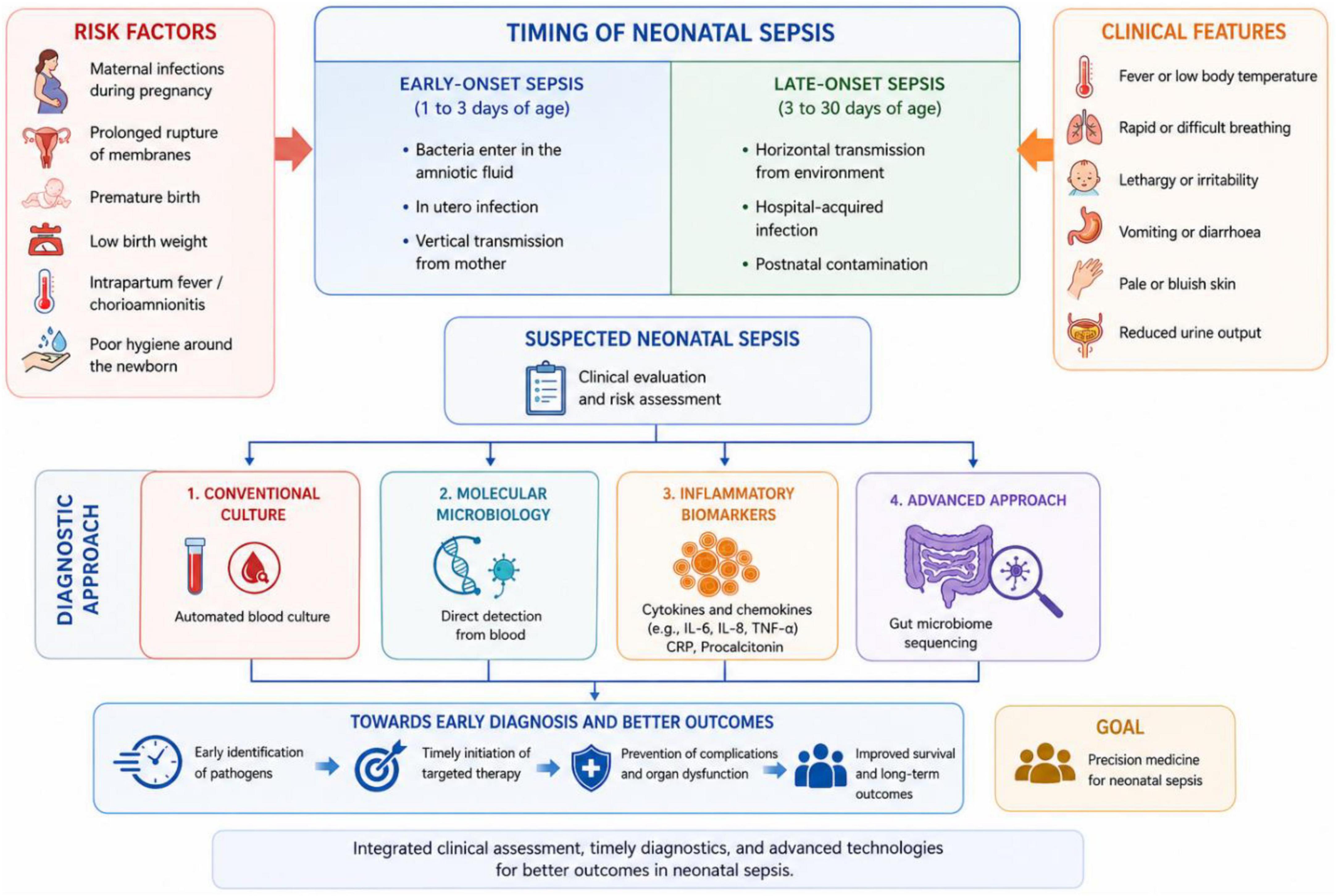
Figure 1. Overview of neonatal sepsis (NS).
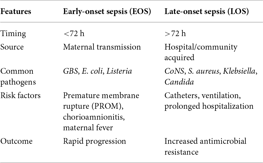
Table 1. Characteristic features of EOS and LOS.
Etiology
The aetiology of NS is influenced by several factors, including the timing of infection onset, duration of hospitalization, prematurity, and the use of invasive procedures such as central venous catheterization. Additional determinants such as the quality of life, cultural practices, and patterns of antibiotic usage also shape the etiological spectrum of sepsis. The common causative agents of NS include a variety of Gram-positive, Gram-negative, anaerobic, and miscellaneous organisms. Among the Gram-positive bacteria, Group B Streptococcus (GBS), Staphylococcus aureus, coagulase-negative Staphylococcus, Enterococcus, and Listeria monocytogenes are predominant. Gram-negative pathogens frequently implicated include Escherichia coli, Citrobacter, Proteus, Klebsiella pneumoniae, Enterobacter, Haemophilus influenzae, Acinetobacter species, and Pseudomonas aeruginosa. Anaerobic organisms such as Bacteroides species, Fusobacterium, and Peptostreptococcus may also contribute to infection. Additionally, atypical pathogens like Chlamydia trachomatis, Mycoplasma, and Ureaplasma are occasionally associated with NS. Congenital neonatal infections are caused by a range of viral, bacterial, and parasitic agents, including Cytomegalovirus, Varicella-zoster virus, Parvovirus B19, Rubella virus, Human Immunodeficiency Virus (HIV), Herpes simplex virus, Plasmodium species, Human T-cell lymphotropic virus type 1, Mycobacterium tuberculosis complex, Toxoplasma gondii, Treponema pallidum, Hepatitis B and C viruses, and Strongyloides stercoralis.
Early onset of sepsis
E. coli and GBS were the most commonly isolated pathogens. With the severity of maternal colonization, neonates are directly associated with the risk of infectious disease. Vernix caseosa or meconium-contaminated amniotic fluid supports growth of GBS and E. coli. Therefore, few species in the vaginal vault will multiply rapidly after premature membrane rupture (PROM), probably contributing to the paradox. Fetal aspiration and ingestion of infected amniotic fluid enables the bacteria to enter the bloodstream and leads to bacteremia (6).
Dissemination of maternal infection occurs in the case of transmission of a few pathogens (e.g. Rubella, Cytomegalovirus, Toxoplasma gondii, Treponema pallidum, L. monocytogenes, and Mycobacterium tuberculosis), which can be transplacental or hematogenic. But most of them are acquired via the ascending infection or when the fetus moves through the infected birth canal during labor (9).
Other agents of EOS include Viridian’s Streptococci, Streptococcus pneumoniae, and Enterococcus species, and Listeria monocytogenes. Enteric gram-negative bacilli like Klebsiella, Citrobacter, and Enterobacter can act as a pathogen. Early-onset S. aureus sepsis is rare but can occur with maternal vaginal colonization and vertical transmission. In premature neonates, nontypeable Haemophilus influenzae sepsis was also reported. Neisseria meningitidis has been reported rarely. Asymptomatic gonorrhea rarely develops during pregnancy, so Neisseria gonorrhoeae can also be a pathogen. The incidence of GBS infection has declined due to the initiation of maternal intrapartum antibiotic prophylaxis in EOS (10).
Late-onset of sepsis
LOS is typically a hospital-acquired infection, although it can rarely be associated with vertical infection. Coagulase-negative Staphylococcus, S. aureus, Enterococcus species, multi-drug-resistant Gram-negative bacilli, and GBS are frequently isolated pathogens (7, 8).
Nearly 70% of Gram-positive organisms, especially CoNS, cause LOS, which is frequently associated with invasive procedures like catheterization. Neonates admitted to the hospital after community exposures are at risk for infections caused by Streptococcus and S. aureus (11).
E. coli is also a major pathogen of LOS in ELBW infants. Other Gram-negative bacilli, such as Klebseilla species, Acinetobacter species, which are multi-drug resistant, are also frequently reported. Outbreaks of Pseudomonas aeruginosa or hospital-acquired sepsis are usually associated with contaminated respiratory equipment (11).
Other pathogens: Anaerobes remain as a causative agent in LOS (especially Bacteroides fragilis). Mortality due to Bacteroides bacteraemia has been documented (12). Candida spp. is a major cause of LOS (12–18%) in ELBW infants. Risk factors of candidemia include abdominal pathology, use of 3rd-generation cephalosporins, hyperalimentation, and central IV catheters (prolonged use—>10 days) (12, 13).
Risk factor of neonatal sepsis
The risk factors for NS are multifactorial, with gestational age and birth weight being the most significant determinants. Risk factors for NS can be broadly categorized into maternal, neonatal, procedural, and environmental factors. Maternal risk factors include chorioamnionitis, PROM, maternal fever, GBS colonization, and urinary tract infections during pregnancy. Neonatal factors such as prematurity, ELBW or LBW, male sex, and low appearance, pulse, grimace, activity, and respiration (APGAR) scores increase susceptibility to infection. Procedural factors, including endotracheal intubation, central venous catheterization, and prolonged antibiotic exposure, further elevate the risk of sepsis, particularly in NICUs. In addition, environmental factors such as poor hygiene practices, contaminated medical equipment, and prolonged hospital stays contribute significantly to the development and transmission of neonatal infections. These factors collectively increase the susceptibility of neonates to infection, particularly those with immature immune systems or prolonged medical interventions (14–16).
Clinical manifestation
Early indications of NS are mostly nonspecific, transient, and indirect. It cannot be distinguished between organisms. They are also indistinguishable from other conditions like respiratory distress syndrome. The variety of symptoms is the result of inflammatory and metabolic processes (17, 18).
Perinatally acquired bacterial sepsis may progress rapidly to death. The initial center of infection can be in the middle ear, genito-urinary tract, paranasal sinuses, gastro-intestinal tract, or lungs. Later it may get disseminated to kidneys, joints, meninges, peritoneum, and bones (19).
Particularly common early signs include anorexia, diminished spontaneous activity, apnea, temperature instability (hypothermia or hyperthermia), bradycardia, somnolence (difficulty in waking the child), poor sucking, poor looking, and poor breathing (18, 20).
Change in body temperature could be a possible indicator of infection. Instability in body temperature (>99.6°F or < 97 °F) can be noticed. Infection is associated with fever or temperature rise greater than 38°C is seen only in 10–15% neonates who develop sepsis (21).
The clinical signs of NS are often nonspecific and may involve multiple organ systems, making early diagnosis challenging. Symptoms are mentioned according to the organ system, and it is given in Table 2.
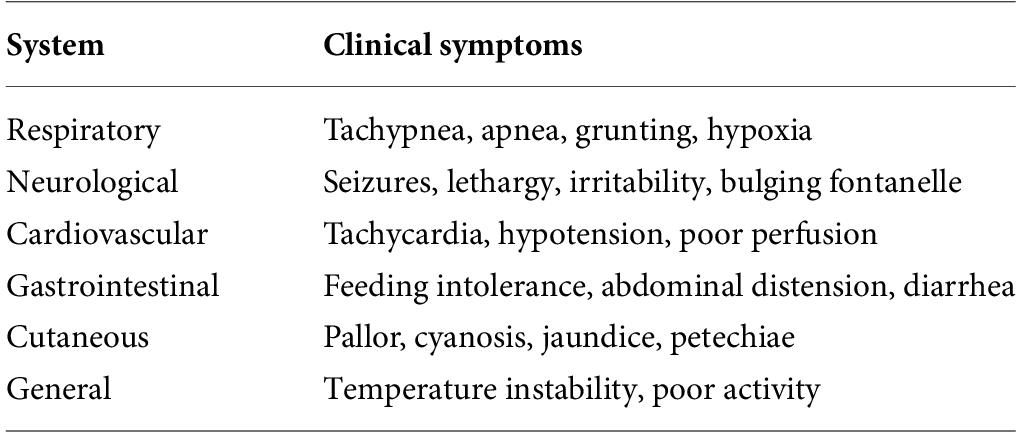
Table 2. Organ system related clinical symptoms.
Many predefined clinical symptoms of infection include the requirement of increased oxygen support or ventilator support and reduced perfusion in the peripherals (capillary refill period >3 sec, increased serum bilirubin, decreased urine production, and metabolic acidosis/base deficiency <−10 mmol/l) (22). This also involves chronic infections such as septicemia, pneumonia, osteomyelitis, meningitis, urinary tract infection, arthritis, and profound diseases (23).
The primary site or a metastatic site of infection can be marked with specific symptoms of an infected tissue.
• Periumbilical erythema, bleeding, or discharge without a hemorrhagic diathesis suggests omphalitis.
• Seizures, a bulging fontanelle, or opisthotonos suggests meningitis, brain abscess, or encephalitis.
• For osteomyelitis or pyogenic arthritis, decreased movement of the extremity and swelling and tenderness over a joint are seen.
• Unexplained abdominal distention may indicate necrotizing enterocolitis or peritonitis (especially when it is accompanied by bloody diarrhea) (24).
Multi-organ failure—heart, lungs, kidneys, and liver—shock, and cerebral edema are the short-term complications. The long-term complications include sensory or neurological dysfunction and delayed developmental milestones (24). Most often sepsis is correlated with congenital heart disease. It can be seen in hemodynamically unstable infants with normal temperatures. It affects up to 0.3% of live births. Early echocardiography is recommended (25).
Systemic inflammatory response syndrome (SIRS)
SIRS can be diagnosed if two of the four parameters are positive (either a or b is mandatory):
a) Abnormal white blood cell count or >10% immature neutrophils
b) Tachypnea >60/min
c) Changes in core temperature
d) Bradycardia or tachycardia (26).
Sepsis was defined as “SIRS in the presence of or as a result of proven or suspected infection.” The causes of sepsis are the following:
• Cytopathology
• Changes in apoptosis rate
• Activation of immunological mediators
• Complement-induced ischemia
Any type of inflammatory stimulus, including infection, leads to the release of cytokines simultaneously. It includes interleukins, which increase the risk of multi-organ failure. It also causes damage to the vessel endothelium and stimulates the coagulation mechanism and increases the activity of fibrinolysis inhibitors (26, 27).
Anti-inflammatory mediators
Mediators trigger the production of acute phase reactants. These are increased in sepsis. After the neonate is stabilized, the sepsis complication is demonstrated by a secondary rise in complete blood count levels. Sepsis can affect neonatal metabolism. This modification may be represented in two phases (27). The first phase is the early shock phase, and the second phase is the flow phase.
Early shock phase
The duration is about 1–3 days. In this phase the entire metabolism of the neonate is slowed. Vasoconstriction, reduced cardiac output, hypoxia, and hypometabolism are the characteristic features of the early shock phase.
Flow phase
In this second phase, the organism enters the hyperactive state. It is triggered by an inflammatory reaction. Mortality is observed in the flow phase. Hyperoxia, increased in cardiac output, hypermetabolism, and increased energy consumption are the characteristic features of the flow phase.
Diagnosis of sepsis
Diagnosis of NS is based on clinical or microbiological evidence. No single laboratory test can effectively diagnose it early stage. Thus laboratory test combinations are used to detect NS (10). Microbiology have a significant role in detecting specific causative organisms. Other modalities for diagnosing NS are based on pathophysiological findings.
Microbiological diagnosis
1. Conventional Blood culture
2. CSF culture
3. Urine culture
4. Biomarkers – CRP, PCT
5. Molecular methods
The possible samples for diagnosing NS include blood, urine, cerebrospinal fluid, umbilical samples like cord blood, and tracheal aspiration (rarely).
Conventional blood culture
Site. Blood can be collected from various sites like the central vein and umbilical catheter. A blood culture specimen should be obtained by sterile vein puncture. Usually blood is collected from a point below the IV line (blood above the IV line usually gets diluted with the fluid being infused).
It can also be done from a newly placed sterile umbilical catheter. Blood cultures may not be accurate from umbilical venous lines, particularly after several hours, as it gets, contaminated by bacteria on the umbilical stump. The specimen should be obtained from the catheter if catheter-associated sepsis is suspected.
Antiseptics. 70% isopropyl alcohol is applied on the skin, followed by an antiseptic solution such as chlorhexidine. Clinicians can also use an iodine tincture and allow the area to dry.
Collection timing. Blood has to be collected before antibiotic therapy, or it can lead to false negative results.
Blood volume. The higher the blood volume, the greater is the yield of isolating pathogens. The minimum volume of blood required is 1.0 ml. It can be difficult to get a sufficient amount of sample from premature infants (11).
Staining. Due to the presence of bacteremia, organisms can be seen in or they can be found in association with polymorphonuclear leukocytes. It can be detected by performing Gram staining, methylene blue staining, or fluorescent staining with acridine orange on the buffy coat.
Blood culture. Identification of a specific pathogen gives several benefits, particularly the ideal antibiotic choice and treatment duration. Blood cultures continue to be the diagnostic gold standard. But it can be difficult to obtain cultures from neonates, as sample amounts are low and a large proportion of samples become contaminated or negative.
Diagnosis is confirmed in blood culture by isolation of a pathogen. For both aerobic and anaerobic organisms, blood should be cultured simultaneously. Pathogens are grown in blood culture bottles using the automated BACTEC™ blood culture system. Conventional methods like plating on media like blood agar and brain heart infusion broth can be used. Growth occurs within 48 h of incubation in >90% of positive bacterial blood cultures.
For candidemia, Candida spp. grows on blood agar plates. Sabouraud’s dextrose agar (SDA) should be used if other fungi are suspected.
Number of blood cultures. It depends on the periodicity of bacteremia. When continuous bacteremia is expected, single blood culture is recommended. In other situations, 2–3 blood cultures are needed to have a good isolation rate (11).
Cerebrospinal fluid culture
Lumbar puncture is only done in neonates who exhibit neurologic signs. Since GBS pneumonia can be mistaken with respiratory distress syndrome that occurs on the first day of life, LP is sometimes performed (12).
Urine culture
Urine culture is required for assessment of sepsis caused by uropathological organisms. Urine should be collected aseptically. The presence of an organism or >5 WBCs in the Gram staining of a urine sample is a probable indication of infection. Absence of pyuria will not exclude it. Culture shows a poor yield, so it is not done routinely (13).
Other possible samples:
1. Umbilical cord blood: EOS is common in preterm infants. So it may be a suitable alternative for improved detection in high-risk neonates. It has increased sensitivity and specificity (14).
2. Tracheal aspiration cultures are beneficial in the first 12 h of life but are not commonly used.
Biomarkers
A biomarker is defined as a “characteristic that can be objectively measured and evaluated as an indicator of normal biological processes, pathological processes, or pharmacological responses to a therapeutic intervention.”
The gold standard for microbiological diagnosis is blood culture, although it has low sensitivity and is a time-consuming procedure. A missing diagnosis causes medication to be delayed and raises the chance of complications. The prognosis of newborn sepsis is determined by early detection and effective antibiotic treatment. As a result, early and precise diagnosis of sepsis is critical for improving clinical outcomes and lowering medical expenses.
Sepsis is a microbial pathogen-induced and a cytokine-mediated disease. After interaction with bacterial proteins, the coagulation cascade and complement cascade get activated and acute-phase reactants are released and serve as a biomarker.
These mediators provide a further opportunity to enhance diagnosis. Efforts have been made over the last decades to improve laboratory diagnosis via the analysis of a wide range of inflammatory biomarkers. Acute phase proteins, pro-inflammatory components like cytokines and chemokines, have been of great interest to detect the progression of NS in recent days.
C-reactive protein (CRP), procalcitonin (PCT), serum amyloid A, interleukin (IL)-6, interleukin-8, soluble triggering receptor expressed on myeloid cells-1 (sTREM-1), neutrophil CD64, and tumor necrosis factor-α are the commonly used biomarkers, and their properties are listed in Table 3.
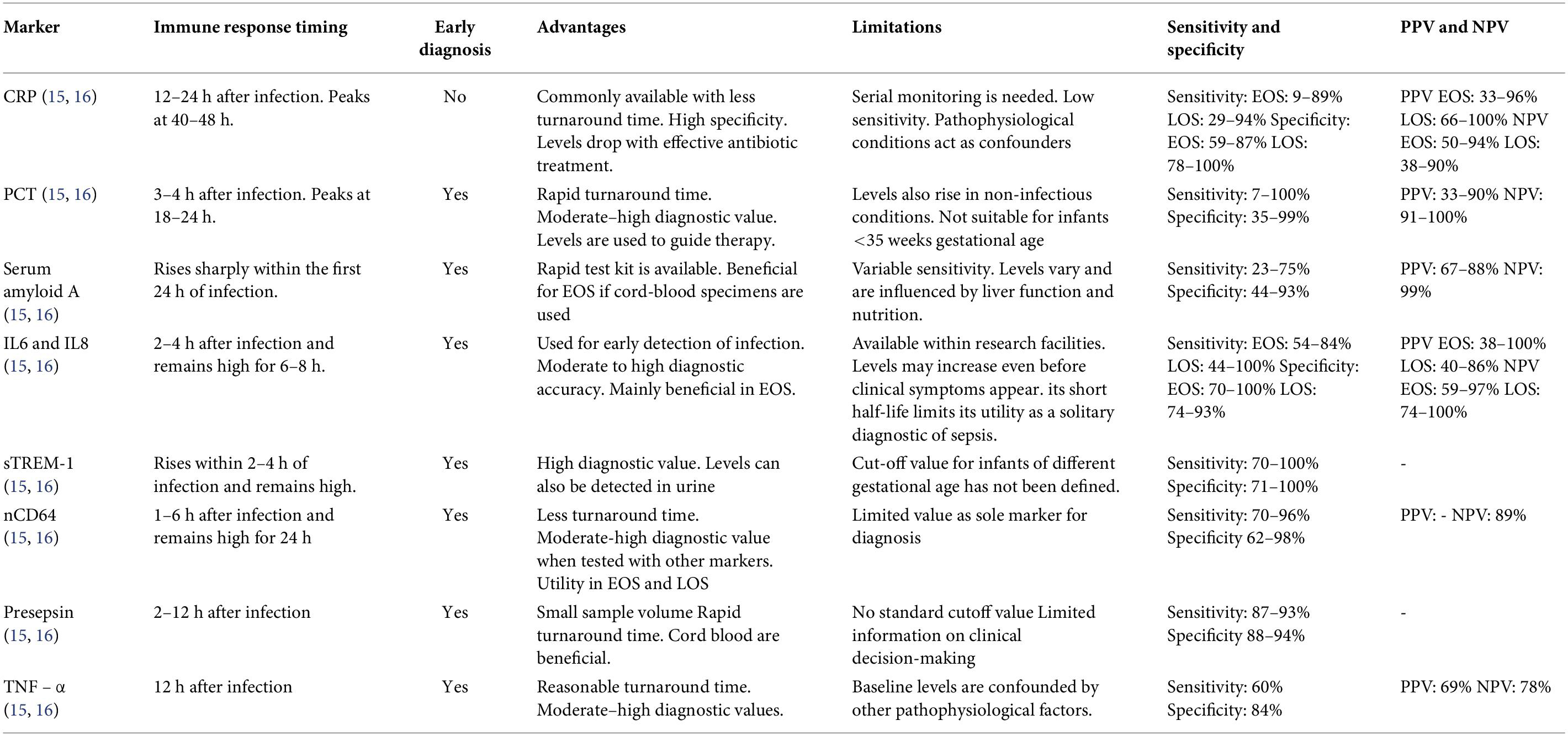
Table 3. Biomarkers in NS.
A good diagnostic biomarker must possess the following clinical properties.
1. It should have a definite cut-off value, high sensitivity, and negative predictive value for “ruling out” disease.
2. Early detection and identifying the category of pathogens.
3. Evaluate disease progression and guide antibiotic use.
4. Predict the disease severity and prognosis.
Currently, no markers are sensitive and accurate to guide the antimicrobial treatment decision regardless of the medical findings. Some cell surface markers provide remarkable outcomes in the experimental framework, but their use is limited in day-to-day clinical practice by the need for specialized tools and skilled personnel. Attempts to enhance diagnostic efficiency were made by integrating several markers (15, 16).
Molecular methods
A molecular technique like polymerase chain reaction (PCR) is used in the identification of causative organisms from blood samples (within a few hours). It can be used to detect the genetic material (DNA/RNA) of the causative pathogen, and properties like virulence and drug resistance can also be identified. It has many advantages, including high sensitivity high specificity, and can quantify bacterial load to assess the efficacy of treatment. Despite high sensitivity and rapid turnaround time, PCR-based methods are associated with several limitations, including contamination-related false-positive results, inability to distinguish viable from non-viable pathogens, restricted pathogen panels, high costs, and limited applicability in resource-constrained settings.
Other diagnosis
a. Biochemistry Analysis
b. Pathological findings with cell markers
c. Role of electrocardiogram and imaging
Biochemistry analysis
1. Blood Glucose measurement – Treat Hypoglycemia
2. Measurement of creatinine and blood urea nitrogen – basic metabolic panel for acute renal failure.
3. Lactic Acid test – Marker for septic shock and severity.
4. Arterial Blood Gas (or Venous Blood Gas) – to detect hypoxia (pO2 < 50 mm Hg) and metabolic acidosis (pH < 7.3, decreased pCO2)
5. Liver Function Tests – It is done if sepsis-associated cholestasis is suspected. Elevated liver enzymes and abnormal serum bilirubin levels are seen in NS (9).
Pathological findings with cell markers
1. CBC
• WBC Count (Decreased < 5000/mm3 or Increased > 25,000/mm3)
• Absolute Neutrophil Count <1000/mm3
• Immature to total neutrophil ratio >0.2
2. Differential WBC count: Neutropenia is a good indicator of sepsis than neutrophilia. Values after 6 h of life are more clinically beneficial than those obtained immediately after birth. The predictive value is improved when the ratio of immature to total neutrophils is >0.2. It has been useful as an early indicator of many infectious conditions, including NS.
3. Platelet count: Thrombocytopenia is also observed and is followed by other Disseminated Intravascular Coagulation (DIC) results. But it is not useful in assessing an NS (18).
Role of electrocardiogram and imaging
Tachycardia and cardiac auscultation findings are the indications for ECG (19). A chest X-ray, and CT head may also be useful in case of pneumonia and subdural hematoma (20).
Association of gut microbiome with neonatal sepsis
The gut microbiome comprises the diverse community of microorganisms that colonize the gastrointestinal tract shortly after birth. These microorganisms play a vital role in neonatal health by promoting immune system maturation, supporting nutrient metabolism, and maintaining the integrity of the intestinal barrier. The composition of the gut microbiome during early life is influenced by factors such as gestational age, mode of delivery, feeding practices, antibiotic exposure, and environmental conditions (Figure 2).
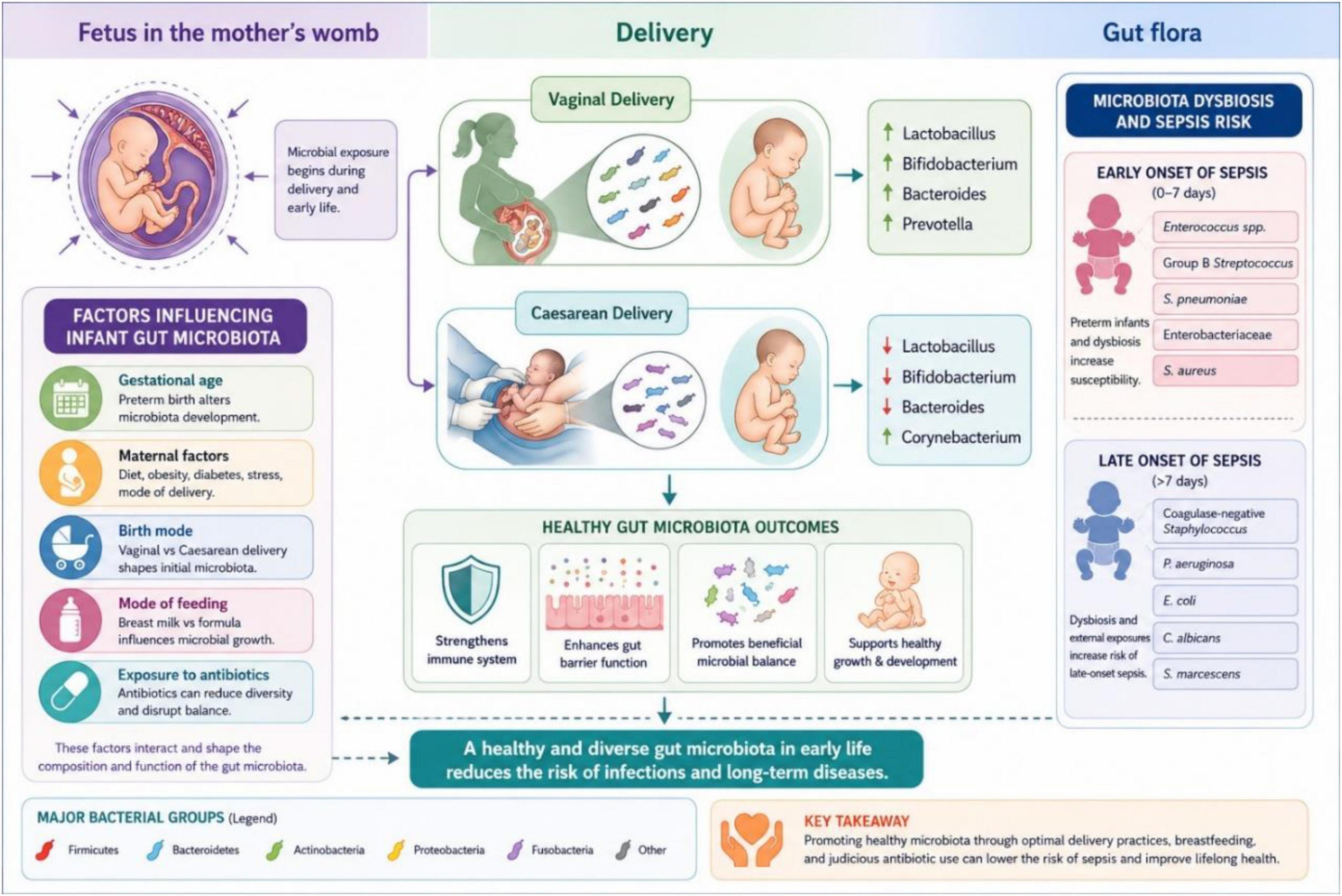
Figure 2. Schematic representation of association of gut microbiome with NS.
In healthy newborns, especially those born vaginally and exclusively breastfed, the gut is predominantly colonized by beneficial bacteria such as Bifidobacterium, Bacteroides, and Lactobacillus. These organisms contribute to host defense by preventing the growth of pathogenic bacteria, enhancing mucosal immunity, and strengthening intestinal barrier function. A balanced microbial community is therefore essential for protecting neonates against infections.
Preterm infants often develop a different microbial profile compared to term infants. Their gut microbiome is typically less diverse and contains lower numbers of beneficial bacteria, while opportunistic pathogens such as Klebsiella, Enterobacter, E. coli, and Enterococcus may become predominant. Factors including prolonged hospitalization, intensive care exposure, invasive procedures, and frequent antibiotic use further disrupt the normal microbial balance, leading to a condition known as gut dysbiosis.
Gut dysbiosis has been increasingly implicated in the development of NS, particularly LOS. An altered microbial environment can compromise the intestinal barrier, allowing bacteria and their products to cross the gut epithelium and enter the bloodstream. This process, known as bacterial translocation, may trigger systemic infection and inflammatory responses. Studies have demonstrated that neonates who subsequently develop sepsis often exhibit reduced microbial diversity and significant alterations in gut microbial composition before the onset of clinical symptoms.
Recent advances in molecular techniques, including next-generation sequencing, have improved understanding of the relationship between the gut microbiome and NS. These technologies have revealed specific microbial patterns associated with increased susceptibility to infection and may facilitate early identification of high-risk infants. Furthermore, the gut microbiome is being explored as a potential therapeutic target. Interventions such as probiotics, prebiotics, and optimized feeding strategies may help restore microbial balance, enhance gut barrier function, and reduce the incidence of NS.
Overall, the gut microbiome plays a central role in neonatal immune development and protection against infection. Disturbances in its composition and function can contribute significantly to the pathogenesis of NS, highlighting its importance as both a biomarker and a target for preventive interventions (21–23).
Management
NS management includes stabilization of a septic newborn and antibiotic therapy. Stabilization can be done, and it includes
• Newborn Resuscitation
• Endotracheal Intubation
• Oxygenation
• Administration Of Intravenous Fluids,
• Administration Of Vasopressors, Glucocorticoids,
• Body Temperature Assessment
• Seizure Management
Antibiotic therapy
Antimicrobial therapy should be initiated immediately in all neonates with suspected sepsis after obtaining appropriate culture specimens, irrespective of initial laboratory findings. Since the early clinical manifestations of NS are often subtle and nonspecific, empirical antibiotic treatment is commonly started while awaiting culture results. Asymptomatic infants with significant perinatal risk factors should also be evaluated carefully and treated when indicated. Due to the increasing prevalence of antimicrobial resistance, empirical monotherapy is generally discouraged.
For early-onset NS, the combination of ampicillin and gentamicin remains the preferred first-line empirical regimen because of its effectiveness against the most common causative organisms. In LOS, antibiotic selection should be guided by local pathogen profiles, institutional antibiograms, and prevailing resistance patterns. Dosages must be individualized according to gestational age, postnatal age, birth weight, and renal function. In clinically stable infants with negative culture results and low suspicion of infection, antibiotics can usually be discontinued after 36–72 h. The duration of treatment typically ranges from 7 to 10 days for uncomplicated bloodstream infections, whereas neonatal meningitis generally requires prolonged therapy of 14–21 days or longer, depending on the pathogen and clinical response (24–26).
Antibiotics used for EOS and LOS are given in Tables 4 and 5.

Table 4. Use of antibiotics for EOS.

Table 5. Use of antibiotics for LOS.
In severe beta-lactam allergy, clindamycin and erythromycin can be considered. If P. aeruginosa is prevalent, ceftazidime, cefepime, or piperacillin/tazobactam may be used. In certain conditions associated with neutropenia, intravenous immunoglobulin may be beneficial. In symptomatic newborns and in blood culture-positive neonates, antibiotics can be continued up to 14 days. Antibiotics can be discontinued if culture is negative after 72 h.
Prevention of NS
• Use of clean delivery kits is mandatory to decrease global incidence. Chlorhexidine application can be done to prevent NS.
Prophylaxis: Universal screening and intrapartum antibiotic use have reduced the incidence of GBS EOS in infants.
If maternal GBS prophylaxis was indicated and provided sufficiently (i.e., penicillin, ampicillin, or cefazolin given IV for ≥4 h), infants should be monitored for 48 h. Diagnosis and medications are done only if symptoms develop.
If sufficient GBS prophylaxis was not given, infants are monitored in the hospital for 48 h. Assessment of risk factors with differential WBC count and CRP levels is done. The clinical course and laboratory findings guide the further management (27).
Role of material science in preventing neonatal sepsis
Advances in material science have introduced innovative strategies for the prevention of NS, particularly in NICUs where invasive medical devices are frequently used. Medical devices such as catheters, feeding tubes, and endotracheal tubes can facilitate microbial colonization and biofilm formation, increasing the risk of infection. To address this challenge, antimicrobial materials and surface coatings containing agents such as silver, chlorhexidine, antimicrobial peptides, and antibiotic-embedded polymers have been developed to inhibit bacterial adhesion and growth. In addition, nanotechnology-based approaches, including silver, zinc oxide, and copper nanoparticles, exhibit broad-spectrum antimicrobial activity through multiple mechanisms such as disruption of microbial cell membranes and inhibition of cellular functions. Emerging smart biomaterials capable of controlled antimicrobial release may further reduce device-associated infections and limit excessive antibiotic exposure. Although many of these technologies are still under evaluation, they represent promising adjunctive measures for reducing the incidence of NS and combating antimicrobial resistance (28, 29).
Conclusion
NS continues to be a major contributor to neonatal morbidity and mortality worldwide, particularly in low- and middle-income countries where healthcare resources remain limited. Despite advances in neonatal intensive care and infection control practices, early diagnosis and timely management remain challenging because clinical manifestations are often nonspecific and laboratory confirmation may be delayed. The increasing prevalence of multidrug-resistant organisms further complicates treatment strategies and underscores the need for judicious antimicrobial use.
Recent developments in diagnostic technologies offer promising opportunities for improving the early detection of NS. In addition to conventional blood culture methods, the integration of molecular techniques, rapid pathogen identification platforms, and biomarker-based approaches may facilitate earlier diagnosis and targeted therapy. Emerging evidence also highlights the important role of the gut microbiome in neonatal immune development and susceptibility to infection. Alterations in microbial composition have been associated with an increased risk of LOS, suggesting that microbiome-based interventions, including probiotics, prebiotics, and optimized nutritional strategies, may contribute to future preventive approaches.
Antimicrobial stewardship has become an essential component of NS management. Rational antibiotic prescribing, timely de-escalation based on microbiological findings, and continuous surveillance of local resistance patterns are necessary to minimize unnecessary antibiotic exposure and reduce the emergence of resistant pathogens. Furthermore, advances in genomics, proteomics, metabolomics, and artificial intelligence are paving the way for precision medicine approaches that may enable individualized risk assessment, early prediction of sepsis, and personalized therapeutic strategies.
Future research should focus on validating novel biomarkers, improving rapid diagnostic tools, elucidating host–microbiome interactions, and developing effective preventive interventions that are applicable across diverse healthcare settings. Strengthening infection prevention practices, enhancing neonatal care infrastructure, and promoting global collaboration in surveillance and research will be critical for reducing the burden of NS. A comprehensive approach that combines early diagnosis, evidence-based treatment, antimicrobial stewardship, and innovative preventive strategies has the potential to significantly improve neonatal survival and long-term health outcomes worldwide.
Funding
The authors declare that financial support was not received for this work and/or its publication.
Conflict of interest
The authors declare that the research was conducted in the absence of any commercial or financial relationships that could be construed as a potential conflict of interest.
References
3. United Nations Inter-Agency Group for Child Mortality Estimation (UN IGME).Levels and Trends in Child Mortality 2024: Report 2024. New York: United Nations Children’s Fund (2024).
5. Desai AP, Leonard EG. Infections in the neonate. In: Fanaroff AA, Fanaroff JM editors. Klaus and Fanaroff’s Care of the High-Risk Neonate. Elsevier (2019). 275 p.
6. Glaser MA, Hughes LM, Jnah A, Newberry D, Harris-Haman PA. Neonatal sepsis: a review of pathophysiology and current management strategies. Adv Neonatal Care. (2021) 21(1):49–60.
7. Fenta GM, Woldemariam HK, Metaferia Y, Seid A, Gebretsadik D. Admission outcome and antimicrobial resistance pattern of bacterial isolates among neonates with suspected sepsis in neonatal intensive care unit at Dessie comprehensive specialized hospital, Dessie, Northeastern Ethiopia. Interdiscip Perspect Infect Dis. (2022) 2022:1318295. doi: 10.1155/2022/1318295
8. Kapourchali FR, Cresci GA. Early-life gut microbiome—the importance of maternal and infant factors in its establishment. Nutr Clin Pract. (2020) 35(3):386–405.
9. Hammad E, Zainab MS. Meta-analysis on factors influencing early onset neonatal sepsis. Sch J Appl Sci Res. (2018) 1(8):20–2.
10. Mangioni D, Peri AM, Rossolini GM, Viaggi B, Perno CF, Gori A , et al. Toward rapid sepsis diagnosis and patient stratification: what’s new from microbiology and omics science. J Infect Dis. (2020) 221(7):1039–47.
12. Pan T, Zhu Q, Li P, Hua J, Feng X. Late-onset neonatal sepsis in Suzhou, China. BMC Pediatrics. (2020) 20(1):1–7.
13. Mohamed W, Algameel A, Bassyouni R, Mahmoud AE. Prevalence and predictors of urinary tract infection in full-term and preterm neonates. Egypt Pediat Assoc Gaz. (2020) 68:1–7.
14. Mithal LB, Palac HL, Yogev R, Ernst LM, Mestan KK. Cord blood acute phase reactants predict early onset neonatal sepsis in preterm infants. PLoS One. (2017) 12(1):e0168677.
15. Sharma A, Thakur A, Bhardwaj C, Kler N, Garg P, Singh M , et al. Potential biomarkers for diagnosing neonatal sepsis. Curr Med Res Pract. (2020) 10(1):12–7.
16. Bunduki GK, Adu-Sarkodie Y. The usefulness of C-reactive protein as a biomarker in predicting neonatal sepsis in a sub-Saharan African region. BMC Research Notes. (2020) 13:1–5.
17. Belachew A, Tewabe T. Neonatal sepsis and its association with birth weight and gestational age among admitted neonates in Ethiopia: systematic review and meta-analysis. BMC pediatrics. (2020) 20(1):55.
18. Procianoy RS, Silveira RC. The challenges of neonatal sepsis management. Jornal de pediatria. (2020) 96:80–6.
20. Ratnaparkhi CR, Bayaskar MV, Dhok AP, Bhende V. Utility of Doppler ultrasound in early-onset neonatal sepsis. Indian J Radiol Imaging. (2020) 30(1):52.
21. Turroni F, Milani C, Duranti S, Lugli GA, Bernasconi S, Margolles A , et al. The infant gut microbiome as a microbial organ influencing host well-being. Ital J Pediatr. (2020) 46(1):1–3.
23. Balasubramanian H, Ananthan A, Rao S, Patole S. Probiotics for preterm infants in India–systematic review and meta-analysis of randomized controlled trials. Indian J Pediatr. (2020) 87(10):817–25.
24. Georgakopoulou I, Chasapi SA, Bariamis SE, Varvarigou A, Spraul M, Spyroulias GA. Metabolic changes in early neonatal life: NMR analysis of the neonatal metabolic profile to monitor postnatal metabolic adaptations. Metabolomics. (2020) 16(5):58.
25. Popescu CR, Cavanagh MM, Tembo B, Chiume M, Lufesi N, Goldfarb DM , et al. Neonatal sepsis in low-income countries: epidemiology,diagnosis and prevention. Exp Rev Anti Infect Ther. (2020) 18(5): 443–52.
26. Dudeja S. Neonatal sepsis: treatment of neonatal sepsis in multidrug-resistant (MDR) infections: part 2. Indian J Pediatr. (2020) 87(2):122–4.
27. Carbone F, Montecucco F, Sahebkar A. Current and emerging treatments for neonatal sepsis. Exp Opin Pharmacother. (2020) 21(5):549–56.
28. Jose A, Gizdavic-Nikolaidis M, Swift S. Antimicrobial coatings: reviewing options for healthcare applications. Appl Microbiol. (2023) 3(1):145–74. doi: 10.3390/applmicrobiol3010012
29. Baltogianni M, Giapros V, Kosmeri C. Antibiotic resistance and biofilm infections in the NICUs and methods to combat it. Antibiotics (Basel). (2023) 12(2):352. doi: 10.3390/antibiotics12020352
© The Author(s). 2026 Open Access This article is distributed under the terms of the Creative Commons Attribution 4.0 International License (https://creativecommons.org/licenses/by/4.0/), which permits unrestricted use, distribution, and reproduction in any medium, provided you give appropriate credit to the original author(s) and the source, provide a link to the Creative Commons license, and indicate if changes were made.